• Skip to main content
  • Skip to secondary menu
  • Skip to primary sidebar
  • About Us
  • Terms of Use
  • Privacy Policy
  • Disclaimer
  • Contact Us
  • Sitemap

BDS Notes

BDS notes, Question and Answers

  • Public Health Dentistry
  • Periodontics
  • Pharmacology
  • Pathology
  • Orthodontics
    • Anchorage In Orthodontics
    • Mandibular Growth, Functional Matrix
    • Retention and Relapse
  • General Surgery
    • Cysts: Types, Causes, Symptoms
    • Maxillofacial Fractures, Disorders, and Treatments
    • Lymphatic Disorders
    • Neurological and Facial Disorders
  • Temporal And Infratemporal Regions
    • Spinal and Neuroanatomy
  • Dental Materials
    • Dental Amalgam
Home » Nasopharynx: Definition, Structure And Function

Nasopharynx: Definition, Structure And Function

February 9, 2026 by Kristensmith Taylor Leave a Comment

Nasopharynx: Definition, Structure And Function

Write a short note on the nasopharynx.
Answer.

Nasopharynx is the upper portion of pharynx lying behind the nasal cavities with which it communicates. Internally it is lined by mucous membrane.

Nasopharynx Boundaries

The roof and lateral wall are formed by the body of the sphenoid which forms a continuous sloping surface, and the basilar part of the occipital bone.

Head And Neck Pharynx and palate Nasopharynx

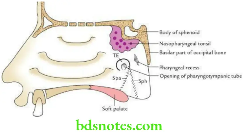
The floor is formed by the sloping upper surface of the soft palate.

Floor Features

  • The presence of the nasopharyngeal tonsil at the junction of the roof and posterior wall, deep to the mucous membrane, is more prominent in children. When enlarged it is called as adenoid.
  • Pharyngeal opening of pharyngotympanic tube which maintains the equilibrium of air pressure on both sides of the tympanic membrane.
  • The tubal tonsil is an aggregation of lymphoid tissue along the upper and posterior margin of the tubal opening deep into mucous membrane producing elevation called tubal elevation.
  • Salpingopharyngeal and salpingopalatine folds. Out of these two folds, one extends downwards towards the wall of pharynx enclosing salpingopharyngeus muscle and is called the salpingopharyngeal fold, while the other extends downwards and forward to the soft palate enclosing the levator palati muscle and is called salpingopalatine fold.
  • The pharyngeal recess is a depression behind the tubal elevation.

Filed Under: Head And Neck

Reader Interactions

Leave a Reply Cancel reply

Your email address will not be published. Required fields are marked *

Primary Sidebar

Recent Posts

  • Branchial Cleft Cyst: Background, Pathophysiology, Etiology
  • Maxillary Nerve: Origin, Course, And Branches
  • The Father Of Anatomy And A Great Anatomist Herophilus
  • Bone Structure – Anatomy
  • The External Carotid Artery: Anatomy, Branches, And Functions
  • Occipitofrontalis Muscle
  • Superficial Temporal Artery
  • Platysma Muscle
  • Cartilage
  • Cauda Equina And Conus Medullaris Syndromes
  • Subcutaneous Injections And Device Management
  • Types Of Circulation: Pulmonary, Systemic, And Portal
  • Structure Of Skeletal Muscle
  • Elastic Cartilage
  • Cellular Organelles And Structure
  • The Golgi Apparatus – The Cell
  • The Cytoplasmic Inclusions Of Certain Plant Cells
  • Dental Abscess
  • Laser Surgery
  • Our Facial Muscles And Their Functions

Copyright © 2026 · Magazine Pro on Genesis Framework · WordPress · Log in