• Skip to main content
  • Skip to secondary menu
  • Skip to primary sidebar
  • About Us
  • Terms of Use
  • Privacy Policy
  • Disclaimer
  • Contact Us
  • Sitemap

BDS Notes

BDS notes, Question and Answers

  • Public Health Dentistry
  • Periodontics
  • Pharmacology
  • Pathology
  • Orthodontics
    • Anchorage In Orthodontics
    • Mandibular Growth, Functional Matrix
    • Retention and Relapse
  • General Surgery
    • Cysts: Types, Causes, Symptoms
    • Maxillofacial Fractures, Disorders, and Treatments
    • Lymphatic Disorders
    • Neurological and Facial Disorders
  • Temporal And Infratemporal Regions
    • Spinal and Neuroanatomy
  • Dental Materials
    • Dental Amalgam
Home » Mucosal Candidiasis Clinical Presentation

Mucosal Candidiasis Clinical Presentation

August 3, 2025 by Joankessler parkland Leave a Comment

Mucosal Candidiasis Clinical Presentation

Question. Write a short note on acute pseudomembranous candidiasis.
Or
Write a short note on oral thrush.

“Understanding the clinical presentation of mucosal candidiasis: Q&A explained”

Answer. It is commonly known as “oral thrush”.

  • Acute pseudomembranous candidiasis appears as a smooth, thick, creamy­white or allow, soft and friable plaque on the oral mucosa.
  • Plaque can be easily wiped of by gentle scraping, which leaves an erythematous, raw, bleeding surface in the underlying area.
  • Lesions may occur at any mucosal site and vary in size ranging from small areas to confluent plaques.
  • Plaque consists of fungal organisms, keratotic debris, inflammatory cells, desquamated epithelial cells, and firin, etc.
  • Oral thrush commonly occurs among children, debilitated elderly persons and AIDS patients.

“Importance of recognizing mucosal candidiasis clinical features: Questions explained”
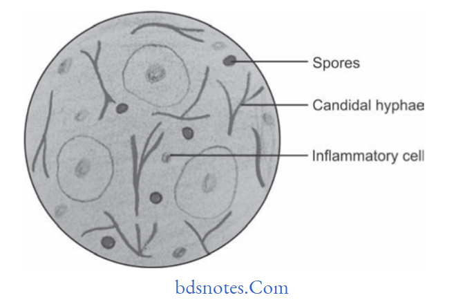
Histopathology

  • Hyperplastic epithelium with superficial necrotic and desquamating parakeratinized layer.
  • Hyperplastic epithelium is infiltrated by candidal hyphae and yeast cells along with PMNs.
  • Often, there is a separation between the superficial pseudomembrane and the deeper layers of epithelium.
  • Candidal hyphae often appear as a weakly basophilic, thread-like structures.
  • Lamina propria is infiltrated by chronic inflammatory cells,i.e., lymphocytes and plasma cells.

“Common challenges in diagnosing mucosal candidiasis effectively: FAQs provided”

Fungal Infections of Oral Cavity Candida albicans

“Steps to explain causes of mucosal candidiasis: Candida albicans vs immune suppression: Q&A guide”

Treatment

  • Anti­fungal drugs, i.e., nystatin, amphotericin­B should be given.
  • Proper oral hygiene should be maintained.

Filed Under: Oral Pathology

Reader Interactions

Leave a Reply Cancel reply

Your email address will not be published. Required fields are marked *

Primary Sidebar

Recent Posts

  • Branchial Cleft Cyst: Background, Pathophysiology, Etiology
  • Maxillary Nerve: Origin, Course, And Branches
  • The Father Of Anatomy And A Great Anatomist Herophilus
  • Bone Structure – Anatomy
  • The External Carotid Artery: Anatomy, Branches, And Functions
  • Occipitofrontalis Muscle
  • Superficial Temporal Artery
  • Platysma Muscle
  • Cartilage
  • Cauda Equina And Conus Medullaris Syndromes
  • Subcutaneous Injections And Device Management
  • Types Of Circulation: Pulmonary, Systemic, And Portal
  • Structure Of Skeletal Muscle
  • Elastic Cartilage
  • Cellular Organelles And Structure
  • The Golgi Apparatus – The Cell
  • The Cytoplasmic Inclusions Of Certain Plant Cells
  • Dental Abscess
  • Laser Surgery
  • Our Facial Muscles And Their Functions

Copyright © 2026 · Magazine Pro on Genesis Framework · WordPress · Log in