• Skip to main content
  • Skip to secondary menu
  • Skip to primary sidebar
  • About Us
  • Terms of Use
  • Privacy Policy
  • Disclaimer
  • Contact Us
  • Sitemap

BDS Notes

BDS notes, Question and Answers

  • Public Health Dentistry
  • Periodontics
  • Pharmacology
  • Pathology
  • Orthodontics
    • Anchorage In Orthodontics
    • Mandibular Growth, Functional Matrix
    • Retention and Relapse
  • General Surgery
    • Cysts: Types, Causes, Symptoms
    • Maxillofacial Fractures, Disorders, and Treatments
    • Lymphatic Disorders
    • Neurological and Facial Disorders
  • Temporal And Infratemporal Regions
    • Spinal and Neuroanatomy
  • Dental Materials
    • Dental Amalgam
Home » Morula

Morula

July 26, 2025 by Kristensmith Taylor Leave a Comment

Morula

Question 1. Meckel’s cartilage.
Answer:

Meckel’s cartilage:

  • It is the cartilage of the first arch.
  • At 6th week of development, this cartilage extends as a solid hyaline cartilaginous rod, surrounded by a fibro cellular capsule, from the developing ear region to the midline of the fused mandibular processes.
  • The two cartilages of each side do not meet at the midline but are separated by a thin band of mesenchyme.
  • It has a close relationship with the development of the trigeminal nerve beginning at two-thirds of the way along the entire length.
  • On the lateral aspect of Meckel’s cartilage, during the sixth week of IU life, a condensation of mesenchyme occurs forming the division of the inferior alveolar nerve.
  • At 7 weeks intramembranous ossification begins in this condensation and then spreads anteriorly up to the middle and posteriorly up to the lingula.

“Factors influencing success with morula studies: Q&A”

  • Remaining Meckel’s cartilage forms.
    • Mental oscillates.
    • Incus and malleus.
    • The spine of the sphenoid.
    • Anterior ligament of mandible.
    • Sphenomandibular ligament.

“Understanding the morula through FAQs: Composition, functions, and uses explained”
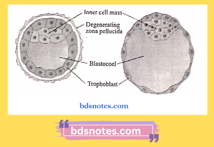
Question 2. Morula.
Answer:

Morula

  • After fertilization of the ovum, a series of cell divisions gives rise to an egg cell mass known as the morula.
  • It looks like mulberry.
  • Fluid seeps into the morula, and the cells realign themselves to form blastocysts,
  • Two cell populations are distinguished within the blastocyst.

“Common challenges in mastering morula notes effectively: FAQs provided”

1. Trophoblast cells.

  • Cells lining, the cavity.
  • It helps to provide nutrition to the embryo.
  • It is associated with the implantation of the embryo and the formation of the placenta.

2. Embryoblast

  • They are inner cell mass.
  • It forms the embryo proper.

“Importance of studying the morula for biology students: Questions explained”

Inner Cell Mass Trophoblast

Filed Under: Anatomy

Reader Interactions

Leave a Reply Cancel reply

Your email address will not be published. Required fields are marked *

Primary Sidebar

Recent Posts

  • Branchial Cleft Cyst: Background, Pathophysiology, Etiology
  • Maxillary Nerve: Origin, Course, And Branches
  • The Father Of Anatomy And A Great Anatomist Herophilus
  • Bone Structure – Anatomy
  • The External Carotid Artery: Anatomy, Branches, And Functions
  • Occipitofrontalis Muscle
  • Superficial Temporal Artery
  • Platysma Muscle
  • Cartilage
  • Cauda Equina And Conus Medullaris Syndromes
  • Subcutaneous Injections And Device Management
  • Types Of Circulation: Pulmonary, Systemic, And Portal
  • Hierarchical Organization Of Skeletal Muscle Tissue
  • Elastic Cartilage Histology Short Note For Medical Exams
  • Cellular Organelles And Structure
  • The Golgi Apparatus – The Cell
  • The Cytoplasmic Inclusions Of Certain Plant Cells
  • Dental Abscess
  • Laser Surgery
  • Our Facial Muscles And Their Functions

Copyright © 2026 · Magazine Pro on Genesis Framework · WordPress · Log in