Midline Diastema
Write a short note on midline diastema.
Or
Write briefly on midline diastema.
Or
Give causes and brief treatment planning on midline diastema.
Or
Give etiology of midline diastema.
Or
Write a short note on the causes of midline diastema.
Or
Give treatment modalities and etiology of midline diastema.
Or
Write short answer on midline diastema and its causes.
Or
Write short note on management of midline diastema.
Or
Write etiology, diagnosis and treatment of midline diastema.
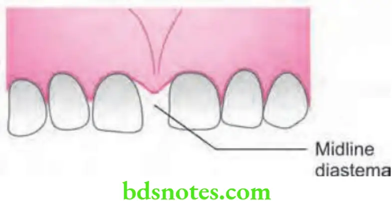
Answer. Midline diastema refers to anterior midline spacing between two central incisors.
It is easy to treat but difficult to retain.
“Understanding the role of midline diastema in dental aesthetics: Q&A explained”
“Importance of studying midline diastema for better orthodontic outcomes: Questions explained”
Causes/Etiology
- Normal/developmental: Midline diastema is present as an incipient malocclusion which gets corrected by itself. Midline spacing occurs at the time of mixed dentition period which is associated with eruption of permanent canines i.e. ugly duckling stage. The condition gets correct by itself as the canine erupts and the pressure transfer from root to coronal area of incisors.
- Tooth material-arch length deficiency: This is a disparity where arch length exceeds the tooth material and leads to midline diastema in conditions such as missing teeth, microdontia, macrognathia and extractions with resultant drifting of adjacent teeth.
- Physical impediment: Presence of a thick flshy labial frenum leads to diastema. This prevents two central incisors from approximating each other as fibrous connective tissue is interposed between them.
“Common challenges in managing midline diastema effectively: FAQs provided”
- Habits
- Abnormal pressure habits such as thumb sucking and tongue thrusting leads to midline diastema. Such patients are present with proclination and generalized anterior spacing.
- Spacing in the midline is caused due to midline soft tissue and hard tissue pathologies such as cysts, tumors and odontomas.
- Presence of an unerupted mesiodens between the roots of two central incisors leads to midline diastema.
- Other causes
- Midline diastema occur during rapid maxillary expansion and it indicates opening of inter-maxillary suture.
- Presence of midline spacing has both racial and familial background. Negroid race show greatest incidence of midline diastema.
“Steps to explain the causes of midline diastema: Genetic vs environmental factors: Q&A guide”
Diagnosis
- First examination and confirmation should be done that whether midline diastema is localized or generalized spacing.
- Assessment of deep overbite should be done.
- Pernicious habits are present or not, this should be checked.
- Blanch test: Upper lip should be lifted and blanching of soft tissues lingual to and between two central incisors should be checked. If blanching is present it means there is high frenal attachment which leads to midline diastema.
- Periapical radiograph: It shows V-shaped notching between central incisors.
- Tooth material arch length deficiency can be determined by model analysis.
Treatment of Midline Diastema
It has three phases i.e.
“Role of frenum attachment in midline diastema formation: Questions answered”
First phase: Identifying and the removal of cause
- This consists of removal of etiology
- Eliminate the habit by use of fied or removable habit breaking appliances.
- Extract the unerupted mesiodens.
- Perform frenectomy
- Any of the midline pathology is treated as indicated.
Second phase: Active treatment
This consists of Active treatment by the use of both removable and the fixed appliances
“Early warning signs of untreated midline diastema: Common questions”
Removable appliances
- Finger springs or a split labial bow can be use to close the midline spacing.
- Provide figer springs distal to two central incisors. A split labial bow can also be used. Labial bow should be extended to the distal aspect of opposite central incisor.
Fixed appliances
- Elastic or springs bring about the most rapid correction of midline diastema.
- Stretch the elastics between two central incisors to close the space. Elastic thread or elastic chain can be used to close the space. An alternative is to stretch the closed coil spring between the two central incisors ‘M’ shaped springs incorporating three helices which are inserted in two central incisor brackets. Activate the spring by closing the helices.
“Asymptomatic vs symptomatic effects of ignoring midline diastema: Q&A”
Third phase: Retention
- This involves the retaining of treated malocclusion.
- Midline diastema is easy to treat and is very difficult to retain.
- Main key behind the successful management is elimination of etiological factors and long term retention by use of certain retainers. Lingual bonded retainers are best for the prolonged retention. Other retainers used are banded retainer’s, hawley’s retainer etc.
“Steps to educate patients about midline diastema and its management: Q&A format”
Other methods of treatment
- Cosmetic restoration: Composites are used to close small midline diastema mainly in adult patients.
- Prosthetic management: An implant or bride is used,
- Surgical management: It is done in median diastema.
- Prosthesis/Crown: Peg-shaped teeth or any other anomalies of shape and size need prosthetic rehabilitation. Replace the missing teeth with fied or removable prosthesis.
- Physiologic diastema which occurs during ugly duckling stage is self correcting condition which requires no treatment.
Leave a Reply