Middle Meningeal Artery
Question 1. Middle meningeal artery
Answer:
It is a branch of the first part of the maxillary artery
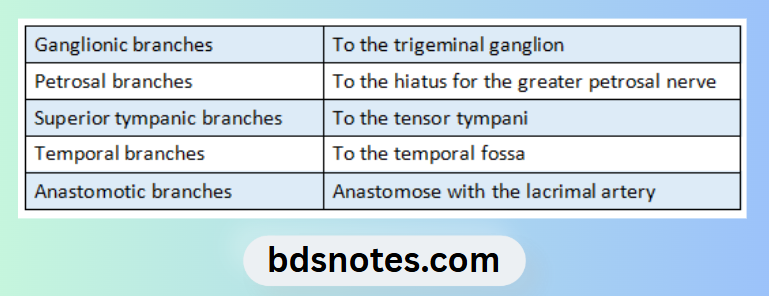
Branches:
“Understanding the middle meningeal artery through FAQs: Anatomy, functions, and uses explained”
“Importance of studying the middle meningeal artery for medical students: Questions explained”
Related Foramen:
- It is transmitted through foramen spinosum
Supplies following structures:
- More of bone & less of meninges
- Bone & red bone marrow in the dipole
- Small branches of duramater
“Role of hematoma in causing epidural bleeding: Questions answered”
Course:
- In the infratemporal fossa, the artery runs upwards & medially deep to the lateral pterygoid muscle & superior to the Sphenomandibular ligament
- It enters the middle cranial fossa through the foramen spinosum
- Here it runs forwards & laterally, grooving the squamous temporal bone & divides into
- Frontal/ Anterior branch:
- It is larger branch
- It runs forwards & laterally towards the lateral end of the lesser wing of the sphenoid
- Parietal/ Posterior branch:
- It runs backwards over, or near the superior temporal sulcus of the cerebrum about 4 cm above the level of the zygomatic arch
Clinical Importance:
It is commonest source of extradural hemorrhage
“Common challenges in mastering middle meningeal artery notes effectively: FAQs provided”
Question 2. Circle of Wills
Answer:
It is an arterial circle, situated at the base of brain in the interpeduncular fossa
Formed by:
1. Anterior & middle cerebral branches of internal carotid artery
- They are interconnected by anterior communicating artery
2. Posterior cerebral branches of basilar artery
- Middle & posterior cerebral artery are united by posterior communicating artery
“Steps to explain disorders affecting the middle meningeal artery: Hematoma vs aneurysm: Q&A guide”
Branches:
- Cortical/ External branches
- Runs on the surface of the cerebrum
- Anastomose freely
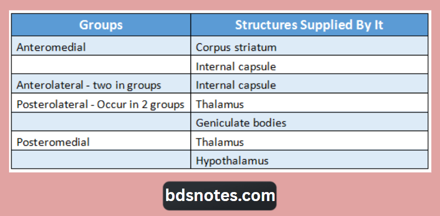
- Central branches
- They are arranged in following groups
“Factors influencing success with middle meningeal artery studies: Q&A”
Leave a Reply