Middle Meatus Of The Nose: Anatomy, Openings, And Clinical Significance
Question 1. Middle meatus of nose
Answer:
- Lies beneath middle concha
- It presents
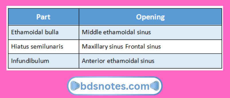
- Ethamoidal bulla
- It is rounded elevation produced by middle ethamoidal sinus
- Hiatus semilunaris
- It is deep semicircular sulcus below bulla
- Infundibulum
- It is short passage at anterior end of hiatus
- Ethamoidal bulla
Middle meatus of nose Openings:
Question 2. Ethmoidal air sinuses
Answer:
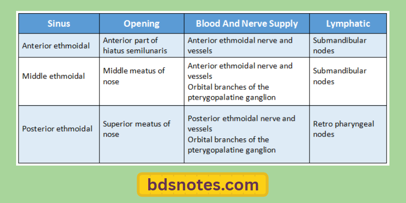
- Ethmoidal sinuses are numerous small inter communicating spaces within the labyrinth of the ethmoid bone
- Bounded above by orbital plate of frontal bone, behind by sphenoidal conchae and the orbital process of the palatine bone and anterior by the lacrimal bone
- Sinuses are divided into anterior, middle and posterior groups
Question 3. Frontal sinus.
Answer:
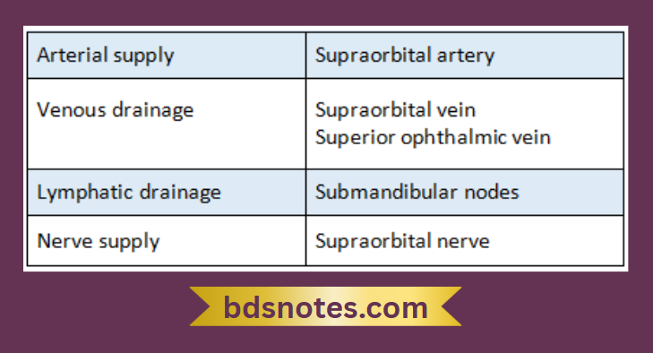
- It lies in the frontal bone deep to the superciliary arch
- It is rudimentary at birth & develops at the age of 7-8 years
Frontal sinus Extend:
- Superiorly- Medial end of the eyebrow
- Posteriorly- Medial part of the roof of the orbit
Leave a Reply