• Skip to main content
  • Skip to secondary menu
  • Skip to primary sidebar
  • About Us
  • Terms of Use
  • Privacy Policy
  • Disclaimer
  • Contact Us
  • Sitemap

BDS Notes

BDS notes, Question and Answers

  • Public Health Dentistry
  • Periodontics
  • Pharmacology
  • Pathology
  • Orthodontics
    • Anchorage In Orthodontics
    • Mandibular Growth, Functional Matrix
    • Retention and Relapse
  • General Surgery
    • Cysts: Types, Causes, Symptoms
    • Maxillofacial Fractures, Disorders, and Treatments
    • Lymphatic Disorders
    • Neurological and Facial Disorders
  • Temporal And Infratemporal Regions
    • Spinal and Neuroanatomy
  • Dental Materials
    • Dental Amalgam
Home » Microscopic Anatomy Of The Kidney: Understanding Renal Pyramids And Lobules

Microscopic Anatomy Of The Kidney: Understanding Renal Pyramids And Lobules

February 6, 2026 by Kristensmith Taylor Leave a Comment

Microscopic Anatomy Of The Kidney: Understanding Renal Pyramids And Lobules

Histology of Kidney
Answer:

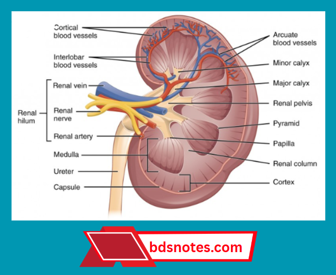
Histology of kidney External structure:

  • It is a bean shaped structure
  • Its convex margin is placed laterally while concave margin is placed medially forming hilum
  • Hilum leads into renal sinus
  • Renal sinus is occupied by the upper expanded part of the ureter called renal pelvis
  • Renal pelvis divides into
    1. Major calyces
    2. Minor calyces
      • Each major calyx divides into it
    3. Papilla
      • It is a projection of kidney tissue

Histology of kidney Internal structure:

  • Kidney consists of
    1. Inner part called medulla
      • It consists of triangular areas called renal pyramid
      • Base of it is directed towards cortex & apex towards renal pelvis
    2. Outer part called cortex
      • It consists of
        1. Cortical arches/lobules
          • It is the tissue lying between the base of the pyramid & surface of the kidney
        2. Renal columns
          • It is the tissue lying between adjacent pyramids

Histology of kidney Lobe of kidney:

  • Pyramid & the cortex around it constitute lobe

Kidney Anatomy Coronal Section

Lymph Node Cellular Components

Filed Under: Anatomy

Reader Interactions

Leave a Reply Cancel reply

Your email address will not be published. Required fields are marked *

Primary Sidebar

Recent Posts

  • Branchial Cleft Cyst: Background, Pathophysiology, Etiology
  • Maxillary Nerve: Origin, Course, And Branches
  • The Father Of Anatomy And A Great Anatomist Herophilus
  • Bone Structure – Anatomy
  • The External Carotid Artery: Anatomy, Branches, And Functions
  • Occipitofrontalis Muscle
  • Superficial Temporal Artery
  • Platysma Muscle
  • Cartilage
  • Cauda Equina And Conus Medullaris Syndromes
  • Subcutaneous Injections And Device Management
  • Types Of Circulation: Pulmonary, Systemic, And Portal
  • Hierarchical Organization Of Skeletal Muscle Tissue
  • Elastic Cartilage Histology Short Note For Medical Exams
  • Cellular Organelles And Structure
  • The Golgi Apparatus – The Cell
  • The Cytoplasmic Inclusions Of Certain Plant Cells
  • Dental Abscess
  • Laser Surgery
  • Our Facial Muscles And Their Functions

Copyright © 2026 · Magazine Pro on Genesis Framework · WordPress · Log in