• Skip to main content
  • Skip to secondary menu
  • Skip to primary sidebar
  • About Us
  • Terms of Use
  • Privacy Policy
  • Disclaimer
  • Contact Us
  • Sitemap

BDS Notes

BDS notes, Question and Answers

  • Public Health Dentistry
  • Periodontics
  • Pharmacology
  • Pathology
  • Orthodontics
    • Anchorage In Orthodontics
    • Mandibular Growth, Functional Matrix
    • Retention and Relapse
  • General Surgery
    • Cysts: Types, Causes, Symptoms
    • Maxillofacial Fractures, Disorders, and Treatments
    • Lymphatic Disorders
    • Neurological and Facial Disorders
  • Temporal And Infratemporal Regions
    • Spinal and Neuroanatomy
  • Dental Materials
    • Dental Amalgam
Home » Median Rhomboid Glossitis

Median Rhomboid Glossitis

July 13, 2025 by Joankessler parkland Leave a Comment

Median Rhomboid Glossitis

Question. Write note on median rhomboid glossitis.
Or
Write short note on median rhomboid glossitis.
Or
Write short answer on median rhomboid glossitis.

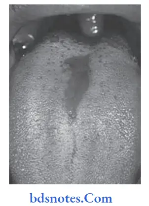
Answer. It is an asymptomatic, elongated, erythematous patch of atrophic mucosa on mid­dorsal surface of tongue.

“Understanding the role of median rhomboid glossitis in oral health: Q&A explained”

Clinical Features

  • Condition is seen mostly in the young adults.
  • It is most common among the males.
  • Lesion is located immediately anterior to the foramen cecum and circumvallate papillae in midline of dorsum of tongue.
  • It starts as narrow, mildly erythematous area located along the median fisure of the tongue.
  • Fully developed lesion of the median rhomboid glossitis appears diamond or lozenge shaped area devoid of papilla.
  • Color of lesion varies from pale pink to bright red.
  • It is usually asymptomatic but occasionally causes slight soreness or burning sensation.

“Importance of studying median rhomboid glossitis for better diagnostic outcomes: Questions explained”

Histopathology

  • Parakeratosis of surface epithelium
  • Loss of papilla
  • Thinning of supra papillary epithelium
  • Presence of acanthosis with elongation of rete ridges.
  • Superficial layer of epithelium shows neutrophilic infiltration and there is presence of candida hyphae.
  • Underlying connective tissue is vascular and is infiltrated by chronic inflammatory cells.
  • The epithelium may show features of dysplasia.

“Common challenges in diagnosing median rhomboid glossitis effectively: FAQs provided”

Developmental Disturbances of Oral and Para oral Structures Median rhomboid glossitis

Filed Under: Oral Pathology

Reader Interactions

Leave a Reply Cancel reply

Your email address will not be published. Required fields are marked *

Primary Sidebar

Recent Posts

  • Branchial Cleft Cyst: Background, Pathophysiology, Etiology
  • Maxillary Nerve: Origin, Course, And Branches
  • The Father Of Anatomy And A Great Anatomist Herophilus
  • Bone Structure – Anatomy
  • The External Carotid Artery: Anatomy, Branches, And Functions
  • Occipitofrontalis Muscle
  • Superficial Temporal Artery
  • Platysma Muscle
  • Cartilage
  • Cauda Equina And Conus Medullaris Syndromes
  • Subcutaneous Injections And Device Management
  • Types Of Circulation: Pulmonary, Systemic, And Portal
  • Structure Of Skeletal Muscle
  • Elastic Cartilage
  • Cellular Organelles And Structure
  • The Golgi Apparatus – The Cell
  • The Cytoplasmic Inclusions Of Certain Plant Cells
  • Dental Abscess
  • Laser Surgery
  • Our Facial Muscles And Their Functions

Copyright © 2026 · Magazine Pro on Genesis Framework · WordPress · Log in