Median Nerve
Question. Describe the median nerve under the following headings: (a) root value, (b) course and relations, (c) branches and distribution, and (d) applied anatomy.
Answer.
The median nerve is so called because it runs in the median plane of the forearm.
“Understanding the median nerve through FAQs: Composition, functions, and uses explained”
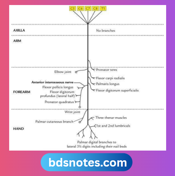
Median Nerve Root value
Ventral rami of C5 to C8 and T1.
“Importance of studying the median nerve for medical students: Questions explained”
Median Nerve Course and Relations
The median nerve is formed in the axilla by two roots – the lateral root from the lateral cord of the brachial plexus and the medial root from the medial cord of the brachial plexus. Then it courses successively through four regions: axilla, arm, forearm, and palm. The medial root crosses the axillary artery to join the lateral root.
Axilla:
In the axilla, the median nerve lies first anterior and then lateral to the axillary artery.
Arm:
In the arm, the median nerve continues to run on the lateral side of brachial artery till the midarm (i.e. insertion of coracobrachialis), where it crosses in front of the brachial artery to lie on its medial side, and then passes anterior to elbow joint to enter the forearm.
“Common challenges in mastering median nerve notes effectively: FAQs provided”
Forearm:
In the forearm, the median nerve passes through the cubital fossa, lying medial to the brachial artery. It leaves the fossa between the two heads of pronator teres before crossing superficial to the ulnar artery from medial to lateral side and giving its anterior interosseous branch below this.
Then it passes deep to the fibrous arch of the flexor digitorum superficialis. It adheres to the deep surface of the flexor digitorum superficialis and leaves the muscle long its lateral border. About 5 cm above the wrist, it lies between the tendons of palmaris longus and flexor carpi radialis. It enters the palm through the carpal tunnel under the flexor retinaculum, but in front of the common synovial sheath enclosing tendons of the flexor digitorum superficialis (FDS) and the flexor digitorum profundus (FDP).
“Factors influencing success with median nerve studies: Q&A”
Palm:
In the palm at the distal border of the flexor retinaculum, it ends by dividing into lateral and medial terminal branches. Before dividing into terminal branches, the median nerve gives off a recurrent muscular branch from its lateral side.
Median Nerve Branches and Distribution
In the axilla:
No branch
In arm:
Muscular branch to pronator teres
“Role of motor innervation in controlling thumb movement: Questions answered”
In the cubital fossa
- Muscular branches to:
- Flexor carpi radialis
- Palmaris longus
- Flexor digitorum superficialis
In the forearm
- Anterior interosseous nerve, which supplies:
- Lateral half of FDP
- Flexor pollicis longus
- Pronator quadratus
- The palmar cutaneous branch supplies the lateral two-thirds of the palm
“Steps to explain functions of the median nerve: Motor innervation vs sensory innervation: Q&A guide”
In the palm
- Recurrent muscular branch, which supplies muscles of the thenar eminence, i.e., abductor pollicis brevis, flexor pollicis brevis, and opponens pollicis
- Lateral terminal branch, which gives off digital nerves to supply both the sides of the thumb and the radial side of the index finger
Note: The digital branch to the lateral side of the index finger also supplies the 1st lumbrical muscle. - The medial terminal branch gives off digital nerves to supply the adjacent sides of the index and middle fingers and the adjacent sides of the index and little fingers the 2nd lumbrical muscle.
Median Nerve Applied Anatomy
The effects of the lesion on the median nerve depend on the site of the lesion.
Effects of Lesions of the Median Nerve

Leave a Reply