• Skip to main content
  • Skip to secondary menu
  • Skip to primary sidebar
  • About Us
  • Terms of Use
  • Privacy Policy
  • Disclaimer
  • Contact Us
  • Sitemap

BDS Notes

BDS notes, Question and Answers

  • Public Health Dentistry
  • Periodontics
  • Pharmacology
  • Pathology
  • Orthodontics
    • Anchorage In Orthodontics
    • Mandibular Growth, Functional Matrix
    • Retention and Relapse
  • General Surgery
    • Cysts: Types, Causes, Symptoms
    • Maxillofacial Fractures, Disorders, and Treatments
    • Lymphatic Disorders
    • Neurological and Facial Disorders
  • Temporal And Infratemporal Regions
    • Spinal and Neuroanatomy
  • Dental Materials
    • Dental Amalgam
Home » Maxillary Sinus: Anatomy And Structure

Maxillary Sinus: Anatomy And Structure

February 9, 2026 by Kristensmith Taylor Leave a Comment

Maxillary Sinus: Anatomy And Structure

Describe the maxillary air sinus under the following headings:

  1. Location,
  2. Boundaries,
  3. Drainage,
  4. Development and
  5. Applied anatomy.

Answer.

Maxillary Air Sinus Location

The maxillary air sinus is the largest paranasal air sinus located in the body of the maxilla.

Maxillary Air Sinus Boundaries

It is a pyramidal-shaped cavity in the body of the maxilla. Its boundaries are as follows:

Head And Neck Nose and paranasal air sinuses Location and relations of maxillary air sinus

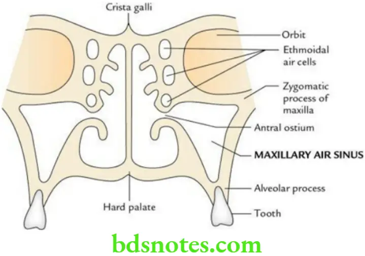
Maxillary Air Sinus Apex:

It is directed towards the zygoma and often extends into the zygomatic bone.

Maxillary Air Sinus Base:

It is formed by the lateral wall of the nasal cavity.

Maxillary Air Sinus Roof:

It is formed by the floor of the orbit.

Maxillary Air Sinus Floor:

It is narrow and formed by the alveolar process of the maxilla. It lies about 1 cm below the level of the floor of the nose.

Maxillary Air Sinus Drainage

It drains in the middle meatus of the nose in the posterior part of the hiatus semilunaris.

Note: The ostium for the maxillary air sinus is located near its roof – a disadvantageous location for natural drainage.

Maxillary Air Sinus Development

It is the 1st paranasal air sinus to develop. It develops in the 4th month of IUL. It grows rapidly during 6–7 years of life and reaches adult size after the eruption of permanent teeth.

Maxillary Air Sinus Applied anatomy

Maxillary Sinusitis (most common):

The maxillary sinus is most commonly infected because its ostium is located near the roof, which hampers its drainage. The infection may reach the sinus either from the nasal cavity or from caries of the upper molar teeth.

Maxillary Sinusitis Referred pain:

The pain of the maxillary sinus may be referred to the upper teeth due to the same nerve supply.

Filed Under: Head And Neck

Reader Interactions

Leave a Reply Cancel reply

Your email address will not be published. Required fields are marked *

Primary Sidebar

Recent Posts

  • Branchial Cleft Cyst: Background, Pathophysiology, Etiology
  • Maxillary Nerve: Origin, Course, And Branches
  • The Father Of Anatomy And A Great Anatomist Herophilus
  • Bone Structure – Anatomy
  • The External Carotid Artery: Anatomy, Branches, And Functions
  • Occipitofrontalis Muscle
  • Superficial Temporal Artery
  • Platysma Muscle
  • Cartilage
  • Cauda Equina And Conus Medullaris Syndromes
  • Subcutaneous Injections And Device Management
  • Types Of Circulation: Pulmonary, Systemic, And Portal
  • Structure Of Skeletal Muscle
  • Elastic Cartilage
  • Cellular Organelles And Structure
  • The Golgi Apparatus – The Cell
  • The Cytoplasmic Inclusions Of Certain Plant Cells
  • Dental Abscess
  • Laser Surgery
  • Our Facial Muscles And Their Functions

Copyright © 2026 · Magazine Pro on Genesis Framework · WordPress · Log in