Maxillary Artery: Branches And Anatomy
Maxillary Artery Origin and extent
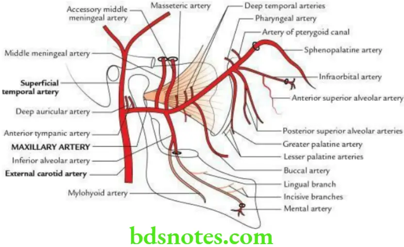
The maxillary artery is the larger of two terminal branches of the external carotid artery. It extends from behind the neck of the mandible to the sphenopalatine foramen, where it continues as the sphenopalatine artery.
“Understanding the anatomy and branches of the maxillary artery through FAQs: Q&A explained”
“Importance of studying the maxillary artery for medical and dental students: Questions explained”
Maxillary Artery Parts
For descriptive purposes, the maxillary artery is divided into three parts by lateral pterygoid (inferior head).
First part: It extends from the neck of the mandible to the point where it crosses the lower border of lateral pterygoid (inferior head).
Second part: It lies superficial or deep to lateral pterygoid.
Third part: It is beyond the upper border of lateral pterygoid. It passes between two heads of lateral pterygoid, passes through pterygomaxillary fissure to enter into pterygopalatine fossa, where it terminates by dividing into sphenopalatine and greater palatine arteries.
“Common challenges in understanding maxillary artery anatomy effectively: FAQs provided”
Maxillary Artery Branches
From first part:
- Anterior tympanic artery
- Deep auricular artery
- Middle meningeal artery
- Accessory meningeal artery
- Inferior alveolar artery
“Factors influencing success with maxillary artery knowledge: Q&A”
From second part: Muscular branches to supply temporalis (deep temporal arteries), pterygoids, masseter and buccinator (buccal branch) muscles.
From third part:
- Posterior superior alveolar arteries
- Greater palatine artery
- Infraorbital artery
- Pharyngeal branch
- Artery of pterygoid canal
- Sphenopalatine artery (the continuation of maxillary artery)
“Steps to explain the anatomy of the maxillary artery: Origin vs course vs branches: Q&A guide”
Maxillary Artery Applied anatomy
Middle meningeal artery: It often ruptures inside the cranial cavity following a trauma on the lateral aspect of the skull and leads to the formation of extradural haematoma.
Inferior alveolar artery: Sometimes it may rupture during extraction of tooth of the lower jaw leading to osteomyelitis of the lower jaw.
Sphenopalatine artery: Its septal branch (rhinologist’s artery) takes part in the formation of Kiesselbach’s plexus in Little’s area of the nose. It is the most common source of nose bleeding.
Leave a Reply