• Skip to main content
  • Skip to secondary menu
  • Skip to primary sidebar
  • About Us
  • Terms of Use
  • Privacy Policy
  • Disclaimer
  • Contact Us
  • Sitemap

BDS Notes

BDS notes, Question and Answers

  • Public Health Dentistry
  • Periodontics
  • Pharmacology
  • Pathology
  • Orthodontics
    • Anchorage In Orthodontics
    • Mandibular Growth, Functional Matrix
    • Retention and Relapse
  • General Surgery
    • Cysts: Types, Causes, Symptoms
    • Maxillofacial Fractures, Disorders, and Treatments
    • Lymphatic Disorders
    • Neurological and Facial Disorders
  • Temporal And Infratemporal Regions
    • Spinal and Neuroanatomy
  • Dental Materials
    • Dental Amalgam
Home » Mastoid Air Cells Of The Temporal Bone: Structure And Function

Mastoid Air Cells Of The Temporal Bone: Structure And Function

February 6, 2026 by Kristensmith Taylor Leave a Comment

Mastoid Air Cells Of The Temporal Bone: Structure And Function

Question 1. Mastoid air cells
Answer:

  • These are series of intercommunicating spaces present within mastoid process

Mastoid air cells Size and number:

  • Varies

Mastoid air cells Blood supply:

Arteries:

  • Posterior tympanic artery from stylomastoid branch of posterior auricular artery

Veins:

  • Mastoid emissary vein
  • Posterior auricular vein
  • Sigmoid sinus

Lymph nodes:

  • Posterior auricular lymph nodes
  • Upper deep cervical lymph nodes

Mastoid air cells Nerves:

  • Tympanic plexus around glossopharyngeal nerve
  • Meningeal branch of mandibular nerve

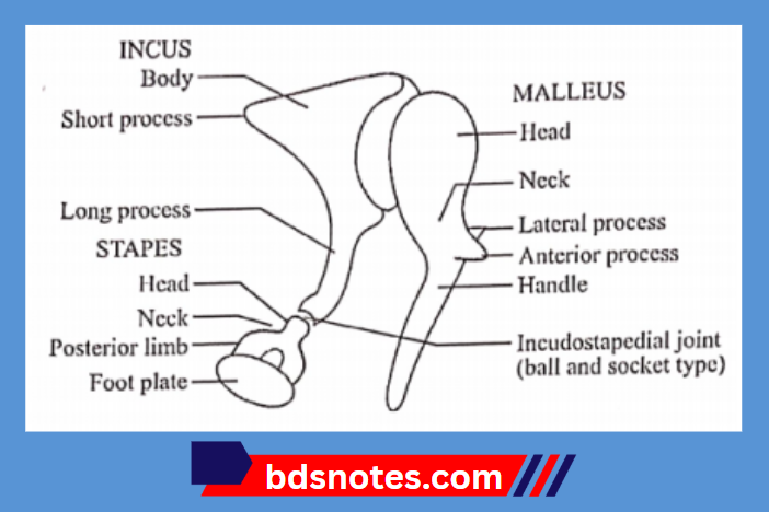
Question 2. Middle ear ossicles
Answer:

1. Malleus:

  • It resembles a hammer
  • It is the largest ossicles

Malleus Parts:

  • Rounded head
  • Neck
  • Anterior process
  • Lateral process
  • Handle

2. Incus or anvil:

  • It resembles an anvil, used by blacksmith
  • It resembles a molar tooth

Incus or anvil Parts:

  • Body
  • Long process

3. Stapes:

  • It is shaped like a stirrup
  • It is smallest, medially placed ossicle

Stapes Parts:

  • Small head
  • Narrow neck
  • Two limbs
  • Footplate, foot-piece or base

Auditory Ossicles Diagram

Filed Under: Anatomy

Reader Interactions

Leave a Reply Cancel reply

Your email address will not be published. Required fields are marked *

Primary Sidebar

Recent Posts

  • Branchial Cleft Cyst: Background, Pathophysiology, Etiology
  • Maxillary Nerve: Origin, Course, And Branches
  • The Father Of Anatomy And A Great Anatomist Herophilus
  • Bone Structure – Anatomy
  • The External Carotid Artery: Anatomy, Branches, And Functions
  • Occipitofrontalis Muscle
  • Superficial Temporal Artery
  • Platysma Muscle
  • Cartilage
  • Cauda Equina And Conus Medullaris Syndromes
  • Subcutaneous Injections And Device Management
  • Types Of Circulation: Pulmonary, Systemic, And Portal
  • Structure Of Skeletal Muscle
  • Elastic Cartilage
  • Cellular Organelles And Structure
  • The Golgi Apparatus – The Cell
  • The Cytoplasmic Inclusions Of Certain Plant Cells
  • Dental Abscess
  • Laser Surgery
  • Our Facial Muscles And Their Functions

Copyright © 2026 · Magazine Pro on Genesis Framework · WordPress · Log in