• Skip to main content
  • Skip to secondary menu
  • Skip to primary sidebar
  • About Us
  • Terms of Use
  • Privacy Policy
  • Disclaimer
  • Contact Us
  • Sitemap

BDS Notes

BDS notes, Question and Answers

  • Public Health Dentistry
  • Periodontics
  • Pharmacology
  • Pathology
  • Orthodontics
    • Anchorage In Orthodontics
    • Mandibular Growth, Functional Matrix
    • Retention and Relapse
  • General Surgery
    • Cysts: Types, Causes, Symptoms
    • Maxillofacial Fractures, Disorders, and Treatments
    • Lymphatic Disorders
    • Neurological and Facial Disorders
  • Temporal And Infratemporal Regions
    • Spinal and Neuroanatomy
  • Dental Materials
    • Dental Amalgam
Home » Mammary Gland Development

Mammary Gland Development

October 27, 2025 by Joankessler parkland Leave a Comment

Mammary Gland Development

Question 1. Describe the development of the breast/mammary gland in brief.
Answer.

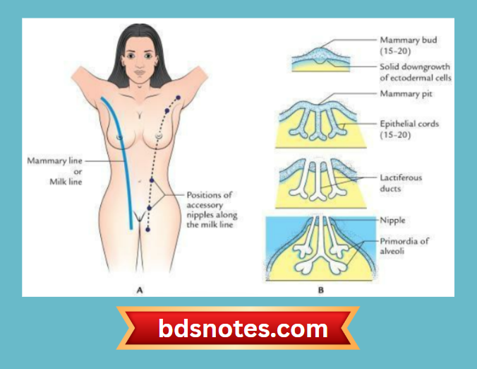
  • The mammary gland develops in the pectoral region from the milk line.
  • The milk line is a linear thickening of surface ectoderm that appears in the 4th week of intrauterine life.
  • The milk line extends from the axilla to the inguinal region on the ventral aspect of the body wall of the embryo.
  • The fibrofatty stroma of the breast develops from the underlying mesoderm.

“Understanding mammary gland development through FAQs: Composition, stages, and uses explained”

Ectodermal Mammary Line Development

“Importance of studying mammary gland development for medical students: Questions explained”

Question 2. Enumerate the congenital anomalies of the breast.
Answer.

  • Polymastia: Supernumerary breasts
  • Amastia: Absence of breast (rare)
  • Athelia: Absence of nipple
  • Polythelia: Supernumerary nipples (commonly seen in the axilla)

“Factors influencing success with mammary gland development studies: Q&A”

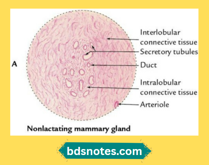
Question 3. Discuss the microscopic/histological structure of the mammary gland.
Answer.

  • The mammary gland is a modified sweat gland of apocrine variety. It is also called a serous, tubuloalveolar gland according to the nature of secretion and secretory units.
  • The histological structure of mammary gland differs according to its physiological status, i.e. (a) nonlactating and (b) lactating.

Microscopic Structure Mammary Gland Ducts Tubules

“Common challenges in mastering mammary gland development notes effectively: FAQs provided”

Microscopic Structure Lactating Breast Tissue

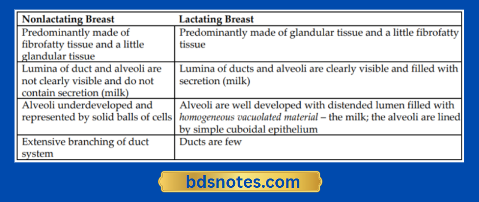
Differences Between Nonlactating and Lactating Breast

Mammary Gland Structure Pre Post Lactation

Filed Under: Upper Limb

Reader Interactions

Leave a Reply Cancel reply

Your email address will not be published. Required fields are marked *

Primary Sidebar

Recent Posts

  • Branchial Cleft Cyst: Background, Pathophysiology, Etiology
  • Maxillary Nerve: Origin, Course, And Branches
  • The Father Of Anatomy And A Great Anatomist Herophilus
  • Bone Structure – Anatomy
  • The External Carotid Artery: Anatomy, Branches, And Functions
  • Occipitofrontalis Muscle
  • Superficial Temporal Artery
  • Platysma Muscle
  • Cartilage
  • Cauda Equina And Conus Medullaris Syndromes
  • Subcutaneous Injections And Device Management
  • Types Of Circulation: Pulmonary, Systemic, And Portal
  • Structure Of Skeletal Muscle
  • Elastic Cartilage
  • Cellular Organelles And Structure
  • The Golgi Apparatus – The Cell
  • The Cytoplasmic Inclusions Of Certain Plant Cells
  • Dental Abscess
  • Laser Surgery
  • Our Facial Muscles And Their Functions

Copyright © 2026 · Magazine Pro on Genesis Framework · WordPress · Log in