Lymphangioma
Lymphangioma is a benign hamartomatous tumor of lymphatic vessels.
Lymphangioma is thought to be developmental malformation of vessels which have poor communication with normal lymphatic system.
“Importance Of Early Detection Of Pyogenic Granuloma”
Lymphangioma Types
Following are the types of lymphangiomas:
- Lymphangioma simplex or capillary lymphangioma: It consists of small thin walled capillaries.
- Cavernous lymphangioma: It consists of dilated lymphatic vessels with surrounding adventitia.
- Cystic lymphangioma or cystic hygroma: It consists of large,macroscopic cystic spaces with surrounding firovascular tissue and smooth muscle.
- Benign lymphangioendothelioma: In this lymphatic channels appear dissecting through dense collagen bundles.
“Risk Factors For Developing Pyogenic Granuloma”
Lymphangioma Clinical Features
- Lymphangioma occur more commonly on head and neck.
- Cervical lymphangiomas are common in posterior triangle and are soft as well as flctuant masses.
lymphangioma
Lymphangioma Oral Manifestations
- Most commonly lesion occurs on tongue and also seen on palate, buccal mucosa, gingiva and lips.
- Superficial lesions occur as papillary lesions which are ofsame color to surrounding mucosa or can be of red hue.
- Deep lesions appear as diffse nodules or masses.
Secondary hemorrhage in lymphatic spaces can cause some of vesicles to appear purple.
“The Role Of Biopsy In Diagnosing Pyogenic Granuloma”
- Tongue involvement results in increase in size of tongue,i.e. macroglossia. Anterior dorsal part of tongue is commonly affcted.
- Lip involvement can lead to macrocheilia.
- Small lymphangiomas are less than 1 cm in size and occur on alveolar ridge of black neonates.
“Early Signs Of Pyogenic Granuloma On The Skin”
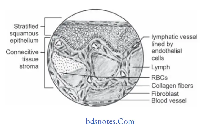
Lymphangioma Histopathology
- Lesion shows multiple, intertwining lymph vessels in loose firovascular stroma.
- Cavernous type show many dilated lymphatics with singlelayer of endothelial cells with flt nuclei and having lymph.
- Vessels which lie beneath surface epithelium replace connective tissue papillae and produce papillary surface change. This appears as translucent vesicle like clinical appearance.
- At times channels can be filed with blood which is known as hemangiolymphangioma.
- At times channels demonstrate proliferation of lymphatic channels with smooth muscle cells known as lymphangiomyoma.
- Cystic hygroma shows cyst like structures.
oral lymphangioma
“Understanding The Role Of Trauma In Causing Pyogenic Granuloma”
“Comprehensive Overview Of Pyogenic Granuloma And Its Significance”
Lymphangioma Treatment
- Surgical removal should be done.
- Complete removal is impossible.
- Surgical debulking is the typical treatment provided.
Leave a Reply