Lipoma: Classification, Pathogenesis, Clinical Features, and Histopathology
Question. Write in brief about lipoma.
Answer. Lipoma is a rare intraoral tumor.
Lipoma is a benign, slow growing tumor of mature fat cells.
“Understanding the role of histopathology in diagnosing lipomas: Q&A explained”
Lipoma Classification
On the basis of morphology
Superficial: It is a single, yellow, lobulated, painless lesion with sessile or pedunculated base.
Diffse: It occurs in deeper tissues and produces a slight surface elevation.
Encapsulated: It is surrounded by a capsule.
Lipoma Pathogenesis
- HMGIC gene which is mapped to 12q15 is a member of high mobility group protein gene family, play a role in development of lipoma.
- Cells of lipoma are different metabolically as compared to normal fat cells. Precursors of fatt acid should be incorporated at faster rate in fat of lipoma as compared to normal fat and there is reduction in lipoprotein lipase activity.
“Importance of studying lipomas for better diagnostic outcomes: Questions explained”
Lipoma Clinical Features
- Lipomas commonly occur in adults, usually around 3rd and 4th decades of life or in older individuals too.
- Lipomas are soft, smooth surface and nodular masses which can be sessile or pedunculated.
- It is asymptomatic for months or years.
- Buccal mucosa and buccal vestibule are the most common sites which are affected. Other sites involved are tongue,flor of mouth and lips.
- The lesion is 3 cm in size but with time, its size increases up to 5 to 6 cm.
“Common challenges in identifying lipomas effectively: FAQs provided”
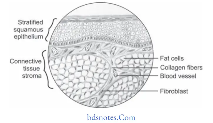
Lipoma Histopathology
“Steps to explain causes of lipoma: Genetic predisposition vs trauma: Q&A guide”
- Lesion consists of mature fat cells and can demonstrate a thin firous capsule.
- Lesion shows the lobular arrangement of fat cells.
- Various microscopic variants of lipoma are appreciated histologically, i.e.
- Fibrolipoma: Fibrous component intermixed with lobules of fat cells.
- Angiolipoma: Admixture of mature adipocytes and multiple small blood vessels.
- Spindle cell lipoma: Variable amount of spindle cell in conjunction with lipomatous component.
- Pleomorphic lipoma: Presence of spindle cells along with bizarre,hyperchromatic giant cells.
- Myolipoma: When spindle cells are of smooth muscle origin
- Intramuscular lipomas: Infitrative growth of mature adipocytes which extend between skeletal muscle bundles.
- Myxoid lipoma: If stromal background is myxoid.
“Role of adipocyte proliferation in causing lipomas: Questions answered”
Lipoma Treatment
Conservative local excision is the treatment of choice.
Leave a Reply