Lichen Planus Causes, Symptoms, and Histopathology
Write short note on lichen planus.
Answer:
“Role of immune dysfunction in causing lichen planus”
Lichen planus is a precancerous condition.
It is a common mucocutaneous disease that arises due to an abnormal immunological reaction and the disease has some tendency to undergo malignant transformation.
Lichen planus
Etiopathogenesis of Lichen Planus
- Oral lichen planus is a T cell-mediated autoimmune disease in which cytotoxic CD8 + T cells trigger the apoptosis of oral epithelial cells. The CD8 + lesional T cells may recognize the antigen associated with major histocompatibility complex (MHC) class 1 on keratinocytes. After antigen recognition and activation, CD8 + cytotoxic T cells may trigger keratinocyte apoptosis. Activated CD8 + T cells may release cytokines that attract additional lymphocytes.
- As per the recent studies in psychoneuroimmunology psychosomatic stress results in autoimmunity reactions and this leads to lichen planus.
“Common causes of lichen planus explained”
Lichen planus Clinical Features
- It occurs among middle-aged and elderly people.
- There is a slight predilection for the females.
- Lichen planus can involve several areas of the oral cavity. Oral lesion: Mucosal surface of buccal mucosa, vestibule, tongue, lips, floor of mouth, palate, and gingiva.
- The patient may report a burning sensation in the oral mucosa.
- The oral lesion is characterized by radiating white and gray velvety thread-like papules in linear, angular, or retiform arrangement; tiny white elevated dots are present at the intersection of white lines known as “Wickham’s striae”.
Lichen planus symptoms
“Importance of early diagnosis of lichen planus”
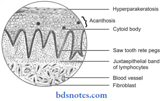
Lichen planus Histopathology
- Overlying surface epithelium exhibits hyper orthokerati nation or hyper para keratinization or both.
- Acanthosis of the spinal cell layer is present.
- Shortened and pointed rete pegs of epithelium produce a “Sawtooth” appearance.
- Intercellular edema in the spinous cell layer is present.
“Techniques for managing oral lichen planus pain”
- There is the presence of necrosis or liquefaction degeneration of the basal cell layer of epithelium.
- Few rounded or ovoid, amorphous eosinophilic bodies are present which are known as “Civatt bodies”.
- These Civatt bodies represent dead keratinocytes or other necrotic epithelial components which are transported to connective tissue for phagocytosis.
- Chronic inflammatory cell infiltration is present in the juxta epithelial lesion.
Lichen planus histopathology
“Impact of stress on triggering lichen planus outbreaks”
Leave a Reply