Question 1. Enumerate the layers of scalp. Give their blood supply, nerve supply & lymphatic drainage. (or) Describe the layers of scalp. Give its nerve supply, blood supply & lymphatic drainage. Add a note on its surgical anatomy. (or) Describe the layers of scalp. Add a note on its applied anatomy (or) Epicranial aponeurosis.
Answer:
- Scalp is the soft tissue which covers the calvaria of the skull
Scalp Layers:
S-Skin:
- S-Skin is outermost layer
- S-Skin is thick and hairy
- S-Skin is adherent to the epicranial
- S-Skin contains large number of hairs, sweat glands, sebaceous gland and is richly supplied by blood vessels
C-Superficial fascia:
- C-Superficial fascia is second layer
- C-Superficial fascia is more fibrous and dense in the center than periphery
- C-Superficial fascia contains large blood vessels & nerves of the scalp
- Thus C-Superficial fascia provides the proper medium for passage of vessels and nerves to the skin
A-Epicranial Aponeurosis:
- A-Epicranial Aponeurosis is freely movable
“Understanding the layers of the scalp through FAQs: Anatomy, functions, and uses explained”
Scalp Attachments:
- Anteriorly: Insertion of frontalis
- Posteriorly: Insertion of occipitalis
- In between occipital bellies: Extrernal occipital protuberance & highest nuchal lines
- Each side: Attached to superior temporal line
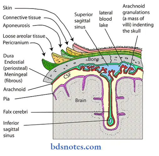
L-Loose areolar tissue:
- L-Loose areolar tissue contains emissary veins devoid of valves
- This communicates the veins of scalp with intracranial venous sinuses
Extent:
- Anteriorly: Eyelids
- Posteriorly: Highest & superior nuchal lines
- Each side: Superior temporal lines
P-Pericranium: Fifth layer
- Loosely attached to bony surfaces
- Firmly adherent to bony sutures
- P-Pericranium: Fifth layer is the outer periosteum of skull Emissary vein
“Importance of studying the layers of the scalp for medical students: Questions explained”
“Common challenges in mastering layers of the scalp notes effectively: FAQs provided”
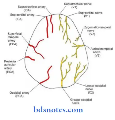
Blood Supply Arterial Supply:

“Why is identifying the layers of the scalp critical for surgical procedures? Answered”
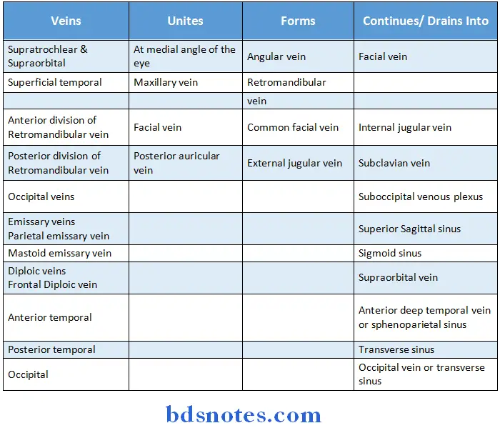
Venous Drainage:
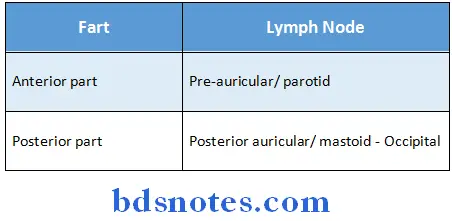
Lymphatic Drainage:
“Factors influencing success with scalp anatomy studies: Q&A”
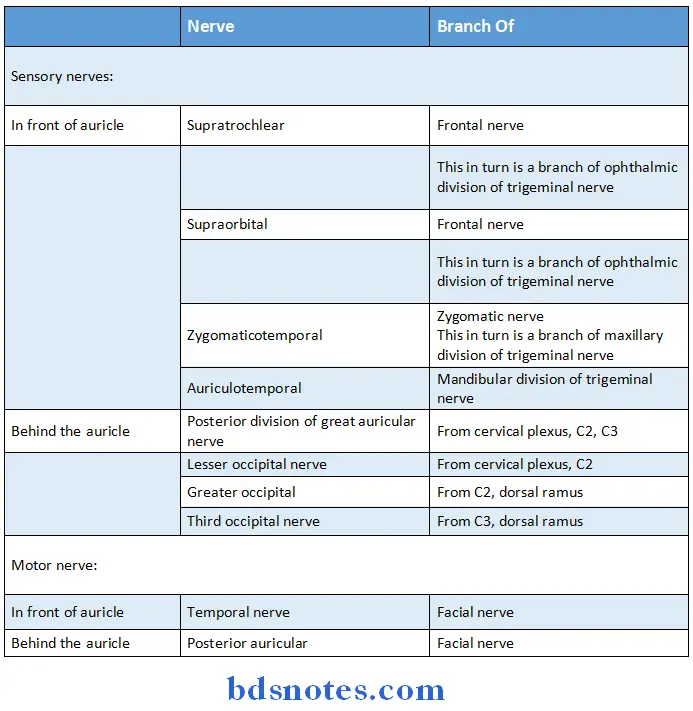
Nerve Supply:
“Steps to explain the layers of the scalp: Skin vs subcutaneous tissue vs galea aponeurotica vs loose connective tissue vs pericranium: Q&A guide”
Applied Anatomy According to layers:
Skin:
- Common site of sebaceous cyst due to abundant sebaceous glands
- Superficial fascia
- Avulsed portion need not be cut away due to rich blood supply
- In open wound the vessels present in this layer are unable to retract & thus produce profuse bleeding
- Inflammation leads to little swelling but much pain
- Loose areolar tissue
- Dangerous area of scalp as emissary vein open here
- If blood is collected in this layer, it leads to generalized swelling
- Epicranial aponeurosis
- Wounds of scalp donot gape unless this layer is divided transversely
- Pericranium
- Collection of fluid deep to it leads to cephalhaematoma
Leave a Reply