Langerhans Cells And Oral Epithelium: Histological Features
Question 1. Langerhans cells.
Answer:
Non-keratinocytes:
Origin: Bone marrow
Site: Suprabasal layer of epithelium.
Structure:
- Dendritic cell
- Has convoluted nucleus
- Has a small rod or flat-shaped granule called a Birbeck granule.
- Lacks desmosomal attachments.
- Appears as a clear cell histologically.
Functions:
- Recognizing and processing antigenic material.
Question 2. Junctional epithelium.
Answer:
- The epithelium of the gingiva which gets attached to the tooth is called the junctional epithelium.
- It resembles reduced enamel epithelium.
- It is non-differentiating, nonkeratinizing tissue
- It is highly permeable and has large intercellular spaces.
- It is formed by the fusion of reduced enamel epithelium and oral epithelium.
- It consists of flattened cells aligned parallel to the tooth surface.
- It has 3-4 layers apically and 15 – 30 layers coronally
Parts:
- Corona part – Thickest and has maximum permeability.
- Middle part – lias maximum adhesion
- Apical part – has maximum mitotic activity.
Question 3. Lamina propria.
Answer:
- The connective tissue supporting the oral epithelium is termed lamina propria.
Layers:
- The superficial papillary layer.
- Associated with the epithelial ridges Collagen fibers are thin and loosely arranged.
- Deeper reticular layer.
- Lies between the papillary layer and the underlying structures.
- Collagen fibers are arranged in thick bundles.
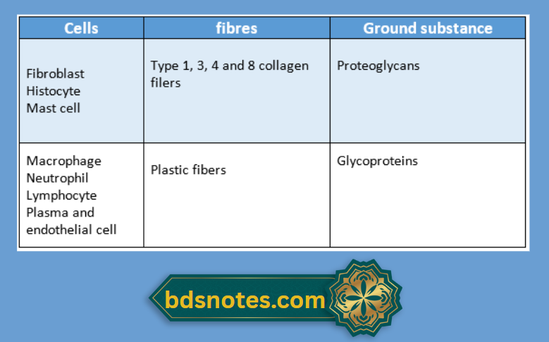
Consist of:
Leave a Reply