• Skip to main content
  • Skip to secondary menu
  • Skip to primary sidebar
  • About Us
  • Terms of Use
  • Privacy Policy
  • Disclaimer
  • Contact Us
  • Sitemap

BDS Notes

BDS notes, Question and Answers

  • Public Health Dentistry
  • Periodontics
  • Pharmacology
  • Pathology
  • Orthodontics
    • Anchorage In Orthodontics
    • Mandibular Growth, Functional Matrix
    • Retention and Relapse
  • General Surgery
    • Cysts: Types, Causes, Symptoms
    • Maxillofacial Fractures, Disorders, and Treatments
    • Lymphatic Disorders
    • Neurological and Facial Disorders
  • Temporal And Infratemporal Regions
    • Spinal and Neuroanatomy
  • Dental Materials
    • Dental Amalgam
Home » Killian-Jamieson Diverticulum: A Rare Cause Of Dysphagia

Killian-Jamieson Diverticulum: A Rare Cause Of Dysphagia

February 9, 2026 by Kristensmith Taylor Leave a Comment

Killian-Jamieson Diverticulum: A Rare Cause Of Dysphagia

Write a short note on Killian’s dehiscence and pharyngeal diverticulum.
Answer.

There is a small, triangular region in the lower part of the posterior wall of the pharynx (the junctional region between the thyropharyngeus and cricopharyngeus), which is not covered by muscles. This weak area is termed Killian’s dehiscence. The mucosa and submucosa of the pharyngeal wall may bulge out through this weak area to form the pharyngeal diverticulum/Zenker’s diverticulum.

Head And Neck Pharynx and palate Pharyngeal diverticulum

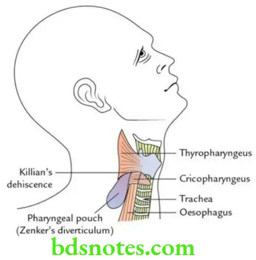
This diverticulum occurs due to neuromuscular incoordination between propulsive thyropharyngeus muscle (supplied by the external laryngeal nerve) and sphincteric cricopharyngeus muscle (supplied by the recurrent laryngeal nerve).

Filed Under: Head And Neck

Reader Interactions

Leave a Reply Cancel reply

Your email address will not be published. Required fields are marked *

Primary Sidebar

Recent Posts

  • Branchial Cleft Cyst: Background, Pathophysiology, Etiology
  • Maxillary Nerve: Origin, Course, And Branches
  • The Father Of Anatomy And A Great Anatomist Herophilus
  • Bone Structure – Anatomy
  • The External Carotid Artery: Anatomy, Branches, And Functions
  • Occipitofrontalis Muscle
  • Superficial Temporal Artery
  • Platysma Muscle
  • Cartilage
  • Cauda Equina And Conus Medullaris Syndromes
  • Subcutaneous Injections And Device Management
  • Types Of Circulation: Pulmonary, Systemic, And Portal
  • Structure Of Skeletal Muscle
  • Elastic Cartilage
  • Cellular Organelles And Structure
  • The Golgi Apparatus – The Cell
  • The Cytoplasmic Inclusions Of Certain Plant Cells
  • Dental Abscess
  • Laser Surgery
  • Our Facial Muscles And Their Functions

Copyright © 2026 · Magazine Pro on Genesis Framework · WordPress · Log in