Key Cells And Landmarks Of The Gingiva: Macrophages & Free Gingival Groove
Question 1. Macrophages.
Answer:
It is cell,present in the lamina propria. n It is a stellate or fusiform cell.
Histology:
- Smaller and denser nuclei
- Less granular endoplasmic reticulum.
- The cytoplasm contains lysosomes and phagocytic vesicles.
Functions:
- Ingest damaged tissue or foreign material.
- Process it and present it to the lymphocytes.
- Stimulates fibroblast proliferation necessary for repair.
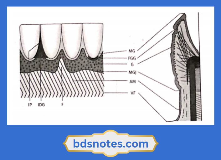
Question 2. Free gingival groove.
Answer:
- It is a dividing line between the free gingiva and the attached gingiva.
- It runs parallel to the margin of the gingiva.
- It is seen as a V-shaped shallow notch.
- It develops at or apical to the bottom of the gingival sulcus.
- Its base is formed by the superior end of the junctional epithelium.
- It is bound on one side by the tooth surface and on the other side by the sulcular epithelium.
Leave a Reply