Keratoacanthoma: Definition, Clinical Features, Histology, and Treatment
Question. Write short note on keratoacanthoma.
Answer. Keratoacanthoma is also known as molluscum sebaceum, selfhealing carcinoma or pseudocarcinoma.
Keratoacanthoma clinically and histologically resembles epidermoid carcinoma so it is mistaken as oral carcinoma.
Keratoacanthoma Etiology
- Hereditary predisposition is present.
- Human papilloma virus (HPV) 26 or 37 can lead to keratoacanthoma.
- Sun exposure
- Chemical agents such as coal tar and minerals
“Understanding the role of histology in diagnosing keratoacanthoma: Q&A explained”
Keratoacanthoma Clinical Features
- Its occurrence is at the age of 50 to 70 years. Male predilection is present.
- Intraorally, it is most commonly found on lips.
- Lesion is painful and regional lymphadenopathy is present.
- Lesion is elevated, umblicated with depressed central core with presence of plug of keratin. Lesion appears as dome shaped.
- Margins of lesions are sharply delineated.
- Lesion begins as small nodule which increases in size from 4 to 6 weeks. Later on it undergoes spontaneous regression from 6 to 8 weeks with scar formation.
“Importance of studying keratoacanthoma for better diagnostic outcomes: Questions explained”
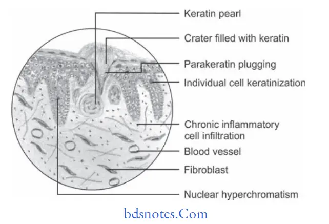
Keratoacanthoma Histopathological Features
“Common challenges in identifying keratoacanthoma effectively: FAQs provided”
- Hyperplastic squamous epithelium growing into underlying connective tissue.
- Epithelial surface is covered by parakeratin or orthokeratin with parakeratin plugging.
- Pseudocarcinomatous infiltration typically present smooth, regular, well demarcated front which does not extend beyond the level of sweat gland.
- Connective tissue show chronic inflmmatory cell infitration.
- Most characteristic feature of lesion is seen at the margins where normal adjacent epithelium is elevated towards central portion of crater, later on an abrupt change in normal epithelium occurs as hyperplastic acanthotic epithelium is reached.
“Asymptomatic vs symptomatic effects of poor communication about keratoacanthoma: Answered”
Keratoacanthoma Treatment
Surgical excision.
Leave a Reply