Keratinized Oral Epithelium: Structure, Layers, And Function
Classify oral mucous and describe keratinized mucosa.
Answer:
Keratinized mucosa:
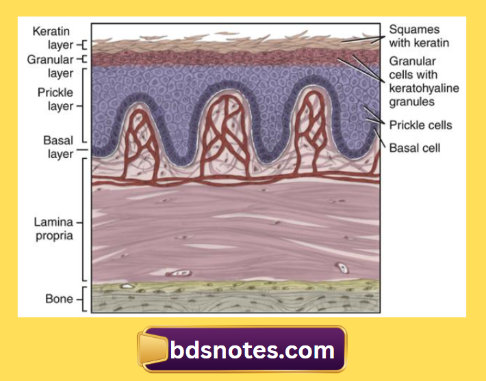
- Keratinizing oral epithelium has keratinocytes arranged in four cell layers.
- Stratum basale
- Stratum spinosum
- Stratum granulosum
- Stratym corneum.
The mucosal surface results from the formation of a surface layer of keratin and the dying process of maturation is called keratinization.
1. Stratum basale:
- It is also called the proliferative or germinative layer.
- It is made up of a single layer of cuboidal cells that synthesize DNA and undergo mitosis to provide new cells.
- They contain bundles of tonofibrils and other cell organelles indicative of protein synthesis.
- The cells in this layer are capable of cell division.
- They divide and form two cell populations.
- One cell population migrate and form cells of the other layer.
- Other cell populations remain as stem cells.
- This layer contains desmosomes – connecting adjacent cells and hemidesmosomes – connecting cells to the basal lamina.
- They provide mechanical linkages.
- Gap junctions allowing electrical and chemical communications are also seen.
2. Stratum spinosum or prickle cell layer:
- Contains several rows of large elliptical or spherical cells.
- Membrane-coating granules appear in the upper part of this layer.
- The nuclei stain less intensely than those of the basal layer.
- They frequently shrink away from each other, remaining in contact only at points known as intercellular bridges.
- This gives the cells a spiny or prickle-like appearance.
- Cells of this layer are the most active in protein synthesis.
- Separation of cells occurs which is caused by loss of intercellular bridges – acantholysis.
- The basal and prickle cell layers together constitute from half to two-thirds of the thickness of the epithelium.
3. Stratum granulosum:
- This layer contains flattened cells containing basophilic keratohyalin granules associated with dense tonofibrils.
- Membrane-coating granules fuse with the cell membrane in the upper part.
- The nuclei of cells show signs of degeneration and pyknosis.
- The cell surfaces become more regular and more closely placed adjacent cell surfaces.
- This layer still synthesizes proteins.
- The membrane-coating granules are glycolipids that have an internal lamellated structure.
- This form an intercellular lamellar material that contributes to the formation of a permeable barrier.
4. Stratum corneum:
- This layer contains extremely flattened and dehydrated cells.
- Cell organelles have been lost.
- Cells are filled only with packed fibrillar material.
- Cells stain bright pink with eosin.
- The keratohyalin granules have disappeared.
- Cells do not synthesize proteins.
Orthokeratinization:
- Cells do not have any nuclei.
Parakeratinization:
- Cells retain pyknotic and condensed nuclei.
- Keratohyalin granules may be present in the cells.
- Cells also contain partially lysed cell organelles until they desquamate.
Leave a Reply