• Skip to main content
  • Skip to secondary menu
  • Skip to primary sidebar
  • About Us
  • Terms of Use
  • Privacy Policy
  • Disclaimer
  • Contact Us
  • Sitemap

BDS Notes

BDS notes, Question and Answers

  • Public Health Dentistry
  • Periodontics
  • Pharmacology
  • Pathology
  • Orthodontics
    • Anchorage In Orthodontics
    • Mandibular Growth, Functional Matrix
    • Retention and Relapse
  • General Surgery
    • Cysts: Types, Causes, Symptoms
    • Maxillofacial Fractures, Disorders, and Treatments
    • Lymphatic Disorders
    • Neurological and Facial Disorders
  • Temporal And Infratemporal Regions
    • Spinal and Neuroanatomy
  • Dental Materials
    • Dental Amalgam
Home » Interpreting An Orthopantomogram

Interpreting An Orthopantomogram

July 28, 2025 by Kristensmith Taylor Leave a Comment

Interpreting An Orthopantomogram

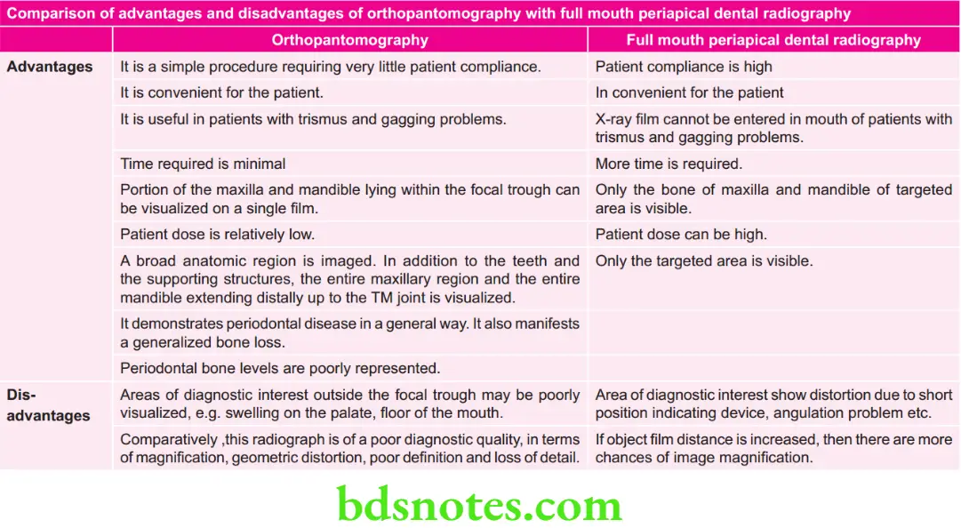
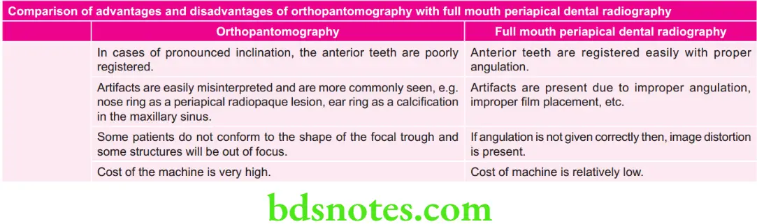
Describe in detail technique of orthopantomography and also write its advantages and disadvantages comparing it with full mouth periapical dental radiography.
Answer.

Synonyms: Panoramic imaging or rotational radiography or pantomography.

Definition: It is a technique for producing a single tomographic image of the facial structures that induces both the maxillary and mandible dental arches and their supporting structures.

Orthopantomography Procedure

  • Explain the procedure to the patient.
  • Make the patient wear a lead apron and remove all objects from the head which will interfere with film exposure, e.g. ear rings, necklace, nose rings.

“Understanding the role of orthopantomograms in dental diagnostics: Q&A explained”

interpreting orthopantomogram

  • Special attntion must be paid for proper positioning of patient along focal trough area.
  • Patient is instructed to look straight rather than following movement of tube.
  • Patient is positioned such that dental arches are located in middle of focal trough area.
  • Mid-sagittl plane is kept perpendicular to flor.

“Importance of studying orthopantomogram interpretation for accurate diagnoses: Questions explained”

  • Patient back and spine is adjusted in erect position and the occlusal plane adjusted so that Frankfurt plane is parallel to flor done by asking the patient to place central incisor into a notched incisal device using a bite block.
  • Mark on fim: Lef and right with lead marker.

Panoramic X-ray interpretation

  • Center the lower border of mandible on the chin rest and is equidistant.
  • Instruct the patient to position the tongue on the palate.
  • After the exposure is complete, then film is subjected to routine processing.

“Common challenges in interpreting orthopantomograms effectively: FAQs provided”

Oral Radiology Panoramic Radiography Or Orthopantography (OPG) Advantages And Disadvantages Of Orthopantomography 1

“Steps to explain different findings on an orthopantomogram: Caries vs periodontal disease: Q&A guide”

Oral Radiology Panoramic Radiography Or Orthopantography (OPG) Advantages And Disadvantages Of Orthopantomography 2

Filed Under: Oral Radiology

Reader Interactions

Leave a Reply Cancel reply

Your email address will not be published. Required fields are marked *

Primary Sidebar

Recent Posts

  • Branchial Cleft Cyst: Background, Pathophysiology, Etiology
  • Maxillary Nerve: Origin, Course, And Branches
  • The Father Of Anatomy And A Great Anatomist Herophilus
  • Bone Structure – Anatomy
  • The External Carotid Artery: Anatomy, Branches, And Functions
  • Occipitofrontalis Muscle
  • Superficial Temporal Artery
  • Platysma Muscle
  • Cartilage
  • Cauda Equina And Conus Medullaris Syndromes
  • Subcutaneous Injections And Device Management
  • Types Of Circulation: Pulmonary, Systemic, And Portal
  • Structure Of Skeletal Muscle
  • Elastic Cartilage
  • Cellular Organelles And Structure
  • The Golgi Apparatus – The Cell
  • The Cytoplasmic Inclusions Of Certain Plant Cells
  • Dental Abscess
  • Laser Surgery
  • Our Facial Muscles And Their Functions

Copyright © 2026 · Magazine Pro on Genesis Framework · WordPress · Log in