Interaction Of X-Ray With Matter
Write in detail about interaction of X-rays with matter.
Answer. As X-rays strike the matter such as tissue of the patient, photons interact with atoms in the absorber and have three possible fates, i.e.
- Coherent scattering (8%)
- Photoelectric effect (30%)
- Compton scattering (62%)
“Understanding the role of X-ray interactions in medical imaging: Q&A explained”
X-rays Coherent scattering
- It is also known as classical or elastic or Thompson scattering.
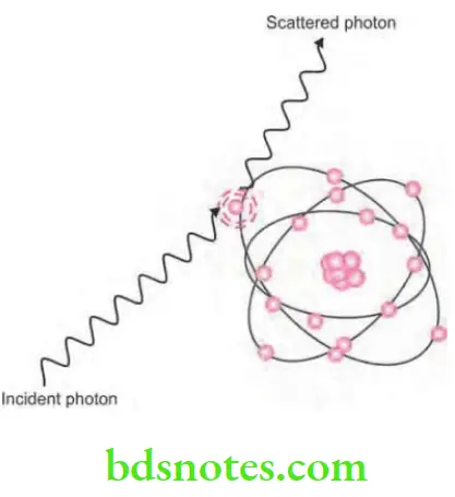
- Coherent scattering is the process by which radiation is deflected without losing energy.
- X-rays when pass close to an atom causes bound electrons to vibrate momentarily at a frequency which is equal to that of incident photon and the incident photon ceases to exist.
- The vibration causes electron to radiate energy in form of another X-ray photon of same frequency and energy as that in the incident beam. Usually, the second photon emitted is at an angle to the path of incidental X-ray.
“Importance of studying X-ray interactions for better diagnostics: Questions explained”
Interaction of X-ray with matter
- This interaction accounts for only 8% of total number of interactions in the dental examination.
- Coherent scattering is negligible in production of fog. This property is used to investigate internal molecular structure of materials by method of X-ray diffraction known as X-ray crystallography.
“Common challenges in explaining X-ray interactions with matter: FAQs provided”
“Steps to explain different types of X-ray interactions: Photoelectric vs Compton effect: Q&A guide”
X-rays Photoelectric Effect
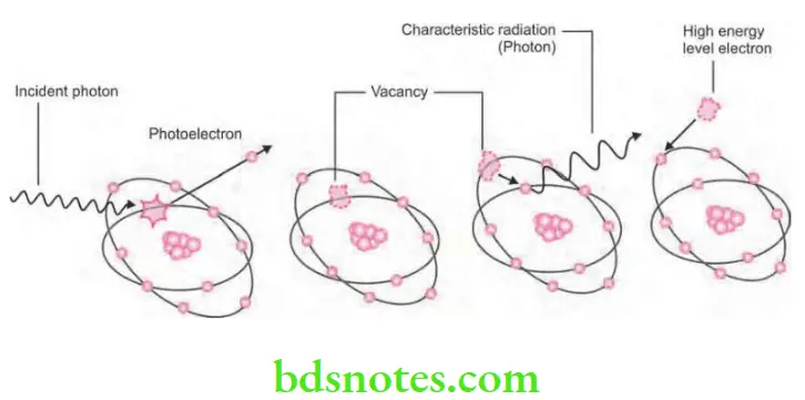
- It is the process of interaction of X-ray photon and the inner shell electron of atom.
- Inner shell electron is ejected which is now known as photoelectron. This will undergo further interactions.
- Energy of the photoelectron is equal to the energy of incident photon minus the ionization energy of inner shell electron.
- Vacancy present in the inner shell is filled by the outer shell electron leading to emission of characteristic radiation. Characteristic photons which are generated are of very low energy that they are absorbed inside the patient and do not fog the film.
Photoelectric effect in radiology
- In this high energy, ejected photoelectrons behaves such as original high energy X-ray photon which undergo many similar interactions and ejecting other electrons as it pass through the tissues.
“Role of photoelectric absorption in X-ray imaging: Questions answered”
“Early warning signs of issues caused by X-ray interactions: Common questions”
- So in the ejected high energy electrons which are responsible for majority of ionization interaction within the tissue and the possible resulting damage which is attributable to X-rays.
- Approximately 30% of photons which are absorbed from dental X-ray beam are absorbed by the photoelectric process.
Radiographic image formation
- In diagnostic radiography, the characteristic radiation generated is of no significance as X-ray photons which are absorbed by the patients are of such a low energy that they are absorbed within the patient. So this is good for dentist as no scattered radiation is present but bad for the patient because of increased radiation absorption.
X-rays Compton scattering
- It is also known as inelastic scattering.
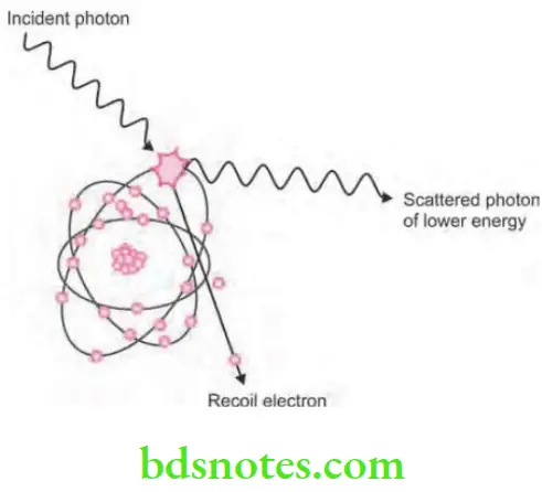
- Compton scattering is the interaction of photons with the free or loosely bound outer shell electrons, i.e. it is an absorption and scattering process predominating with higher energy photons.
- X-ray photon interacts with the outer shell electron of tissue atom and get ejected which is known as Compton recoil electron, with some energy of incoming photon this means there is some absorption.
Coherent scattering in X-ray
- Ejected electron undergoes further ionizing interaction inside the tissues.
- Remainder of incoming photon energy is deflected or scattered from original path as scattered photon.
- Scattered photon then undergo further Compton interaction inside the tissues; photoelectric interaction inside the tissues and escape from the tissues.
- Photons which escaped from the tissues form scatter radiation in clinical era.
- Approximately, 62% of photons are absorbed from dental X-ray beam by this process.
“Steps to educate patients about X-ray interactions and their safety: Q&A format”
“Asymptomatic vs symptomatic effects of ignoring X-ray interaction principles: Q&A”
- The importance of photoelectron and Compton absorption in diagnostic radiography relates to the difference in the way photons are absorbed in various anatomic structures.
X-ray attenuation in the human body
- The probability of both photoelectric and Compton interactions of photons with matter is higher in hard tissues than in less mineralized soft tissues. There are more photons in the beam exiting the patient after traversing soft tissue than through hard tissue. As a consequence, a radiograph readily distinguishes between many tissues, including enamel, dentine, bone, and soft tissue.
Leave a Reply