Hyperplastic Pulpitis
Question. What is pulpitis? Explain the different causes of pulpitis.
Describe clinical features and the histopathological picture of chronic open hyperplastic pulpitis.
Answer.
Pulpitis:
Pulpitis refers to the inflammation of the dental pulp within the tooth.
Causes of pulpitis
- Reversible pulpitis is caused by an agent capable of injuring the pulp, such as trauma, disturbed occlusal relationship, or thermal shock.
- Irreversible pulpitis: It is caused by the bacterial involvement of pulp through caries, chemical or thermal, or mechanical injury.
“Role of chronic irritation in hyperplastic pulpitis”
Chronic open hyperplastic pulpitis
- It is also known as chronic hyperplastic pulpitis or pulp polyp, or pulpitis aperta.
- It is a productive pulpal inflammation due to an extensive carious exposure of the young pulp. It is characterized by the development of granulation tissue covered by epithelium and resulting from long-standing low-grade infection.
pulp polyp treatment
hyperplastic pulpitis
“Clinical examination for hyperplastic pulpitis”
Clinical Features
- Pulp polyp appears as a small, pinkish, red-lobulated mass, which protrudes from pulp chamber and fills up the carious cavity.
- The condition is seen in young adults and children. It commonly develops in deciduous molars and first permanent molars.
- The affected tooth has a large open carious cavity, which has been present for a long duration.
- The lesion bleeds profusely on provocation.
- The involved tooth is painless and is sensitive to thermal stimuli.
“Pathophysiology of hyperplastic pulpitis explained”
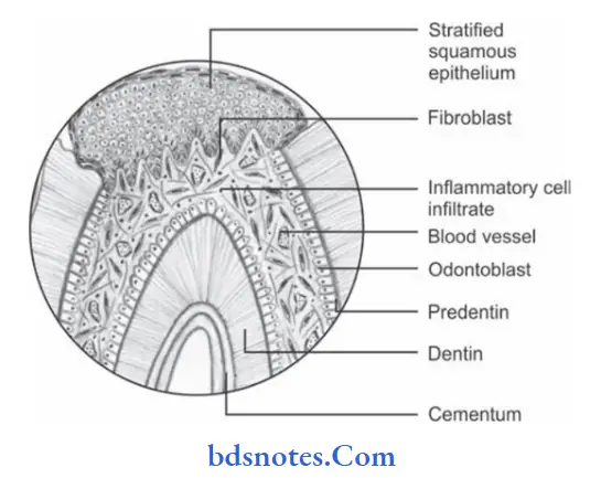
Histopathology
“Root canal therapy for hyperplastic pulpitis”
- Hyperplastic pulp tissue lesion presents the feature of granulation tissue mass, consisting of numerous proliferating fibroblasts and young blood capillaries.
- Inflammatory cell infiltration by lymphocytes, plasma cells, and sometimes polymorphonuclear neutrophils in tissue is common.
- Stratified squamous epithelium is present on the surface of hyperplastic pulpitis, which resembles oral epithelium.
- The epithelial surface shows well-formed rete peg formation.
- The epithelial cells on the surface are believed to be desquamated epithelial cells that came either from the buccal mucosa or salivary gland ducts.
Leave a Reply