How to Identify Dentigerous Cyst on X-ray – Radiographic Guide
Dentigerous Cyst
Write a short note on the dentigerous cyst.
Answer:
Dentigerous cyst
It is also known as a follicular cyst.
Dentigerous cyst Pathogenesis
- Intrafollicular Theory: Dentigerous cyst is caused by a fluid accumulation between reduced enamel epithelium and enamel surface which results in a cyst in which the crown is located within the lumen.
- Extrafollicular Theory: Dentigerous cyst may arise by proliferation and cystic transformation of islands by odontogenic epithelium in a connective tissue wall of a dental follicle or even outside dental follicle and this transformed epithelium then unite with lining follicular epithelium forming cystic cavity around tooth crown.
Dentigerous cyst X-ray
“Importance of early detection of dentigerous cysts on X-rays”
Dentigerous cyst Radiographic Features
A dentigerous cyst reveals a unilocular radiolucent area which is associated with the crown of an unerupted or impacted tooth. There are various radiographic types which are given by Thoma.
- Central Variety: In this, the crown is enveloped symmetrically. The pressure which is applied by the cystic fluid to the crown of the tooth may push the tooth away from its direction of eruption.
- Lateral Variety: In this, the radiographic appearance of the dentigerous cyst occurs due to the dilatation of the follicle on one aspect of the crown. This is associated with mesioangular impacted mandibular third molars which are partially erupted.
- Circumferential Variety: In this, the complete tooth is enveloped by the cyst.
Radiographic features of dentigerous cyst
“Can dentigerous cysts recur after treatment?”
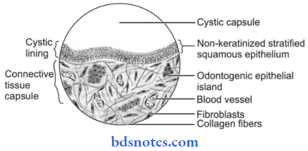
Dentigerous cyst Histologic Features
Non-inflamed Dentigerous Cyst
- H&E stained section shows stratified squamous epithelium which is 2 to 4 cell layer thick and lines the lumen.
- Rete peg formation is absent. So the epithelium connective tissue interface is flat.
- The fibrous connective tissue wall is loosely arranged.
“Treatment options for dentigerous cyst identified on X-rays”
Inflamed Dentigerous Cyst
- H&E stained section shows stratified squamous epithelium which shows varying amounts of hyperplasia.
- Small islands or cords of inactive appearing odontogenic epithelial rests at times present in the fibrous walls.
- Rete peg formation is present.
- Focal areas of mucous cells are also seen in the epithelium of dentigerous cysts.
- Rarely the ciliated columnar cells are seen in the epithelium. Very rarely small nests of sebaceous cells are seen in fibrous cyst walls.
- Rushton bodies are present in epithelium. Rushton bodies are linear, curved, hyaline bodies which show variable stainability and have an uncertain origin.
- The connective tissue wall is thick and consists of loose fibrous connective tissue or sparsely collagenized myxomatous tissue.
- There is the presence of chronic inflammatory cell infiltration which consists of lymphocytes and plasma cells.
- Cystic lumen consists of a thin watery yellow fluid that is occasionally blood-tinged.
Dentigerous cyst radiographic appearance
“Pathophysiology of dentigerous cysts explained”
Leave a Reply