How to Differentiate Dystrophic and Metastatic Calcification
Write A Short Note On Pathological Calcification.
Answer:
Pathologic calcification is the abnormal deposition of calcium in various tumors and organs of the body:
“Techniques for managing symptoms of pathological calcification”
They are of three types:
- Dystrophic calcification
- Metastatic calcifiation
- Calcinosis.
Dystrophic vs metastatic calcification
“Importance of distinguishing between dystrophic and metastatic calcification”
Dystrophic Calcification
- It is a type of pathologic calcification in which calcium salts are deposited in the dead or degenerating tissue of the body.
- It is not associated with increased levels of serum calcium and is related to changes in the local environment.
- This is the most frequent type of pathological calcification found in a wide variety of tissues.
- In the mouth, the area of dystrophic calcification is found in the gingiva, tongue, cheek, and pulp.
- One of the most common intraoral dystrophic calcifications found in the pulp of the teeth is “Pulp Stone.”

“Common causes of pathological calcification explained”
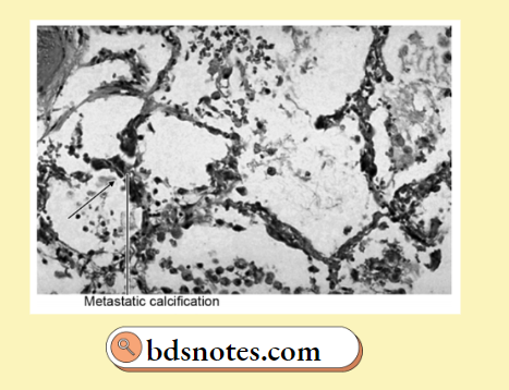
Metastatic Calcifiation
- Abnormal deposition of calcium in the tissue due to an increase in the amount of serum calcium.
- It occurs particularly in diseases like hyperparathyroidism, which depletes bone calcium and causes high levels of blood calcium.
- Metastatic calcification also occurs in hypervitaminosis D. In this type of calcification, a deposit of calcium occurs in the kidney, lung, gastric mucosa, and media of blood vessels.
“Role of tissue damage in causing dystrophic calcification”
Pathological Calcification Calcinosis
- Abnormal deposition of calcium under the skin is also known as calcinosis.
- There are two forms of calcinosis:
- Calcinosis circumscripta: It is circumscribed form.
- Calcinosis universalis: It is a generalized form and is associated with scleroderma and dermatomyositis.
Metastatic calcification
Gross Pathology Of Pathological Calcification
Pathologic calcification appears as fine white granules or clumps of gritty deposits.
“Case studies on outcomes of dystrophic and metastatic calcification”
Microscopical Picture Of Pathological Calcification
- In Hematoxylin and Eosin (H&E) stained sections, it appears as intracellular or extracellular basophilic amorphous granular deposits.
- At times, single necrotic cells act as seeds that get encrusted with lamellar mineral deposits, i.,e. psammoma body is called so due to its resemblance to grains of sand and is commonly seen in some papillary cancers, i.e., thyroid and meningiomas.
- Calcium and iron salts may gather long, slender spicules of asbestos in the lungs, creating beaded, dumbbell forms called asbestos bodies.
“Impact of hypercalcemia on metastatic calcification”

Leave a Reply