Hodgkin’s Lymphoma: Clinical Features, Histopathology, and Treatment Explained
Question. Write in detail about Hodgkin’s lymphoma.
Answer. It is also known as Hodgkin’s disease.
- Epstein-Barr virus is considered to be the major cause.
- Patients with HIV infection have a higher incidence of Hodgkin’s disease.
“Understanding the role of histopathology in diagnosing Hodgkin’s lymphoma: Q&A explained”
Hodgkin’s Lymphoma Clinical Features
- Hodgkin’s Lymphoma is most commonly seen in young adults and older individuals.
- More common in males as compared to females.
- Clinical signs and symptoms of the disease are protean
- There is painless enlargement of one or more cervical lymph nodes.
- Palpable painless cervical lymphadenopathy occurs in cervical area, axilla and less commonly in inguinal area and Waldeyers ring and occipital nodes.
- Lymph nodes are fim and rubbery in consistency and overlying skin is normal.
- Symptoms are of unexplained weight loss, fever and night sweats.
“Importance of studying Hodgkin’s lymphoma for better diagnostic outcomes: Questions explained”
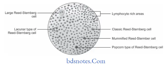
Hodgkin’s Lymphoma Histopathology
- Nodular sclerosis hodgkin’s disease
- Morphology show nodular pattrn.
- Broad bands of fiers divide node into nodules
- Characteristic cell is lacunar type ReedSternberg Cell which has monolobated, multilobated nucleus and a small nucleolus with abundant and pale cytoplasm.
- Mixed cellularity hodgkin’s disease
- Infiltrate is usually diffuse
- ReedSternberg cells are of classic type i.e. large with bilobate, double or multiple nuclei and a large eosinophilic inclusion like nucleolus.
“Common challenges in diagnosing Hodgkin’s lymphoma effectively: FAQs provided”
- Lymphocyte-depleted hodgkin’s disease
- Infiltrate is diffuse and often appears hypocellular.
- Large number of ReedSternberg cells and bizarre sarcomatous variants are present.
- It is associated with older age and HIV positivity
- Lymphocyte-rich classic hodgkin’s disease
- ReedSternberg cells of the classic or lacunar type are observed with background infitrate of lymphocytes.
- Nodular lymphocyte-predominant hodgkin’s disease
- In this typical ReedSternberg Cell is not seen, instead a variant of Reed
- Sternberg Cell the lymphocytic and histiocytic cells or popcorn cells are seen within the background of inflammatory cells which are predominantly benign lymphocytes.
“Steps to explain causes of Hodgkin’s lymphoma: Genetic vs environmental factors: Q&A guide”
“Role of Epstein-Barr virus in causing Hodgkin’s lymphoma: Questions answered”
Hodgkin’s Lymphoma Treatment
Combination of radiotherapy and chemotherapy help in curing the disease.
Leave a Reply