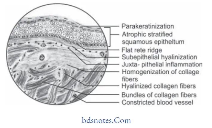
Histopathology Of Oral Submucous Firosis
Following is the common histopathological features of oral submucous fibrosis:
“Importance Of Studying Histopathology In Oral Submucous Fibrosis”
- Overlying hyper keratinized or atrophic epithelium often shows flttning and shortening of rete pegs.
- There can be variable degrees of cellular atypia or epithelial dysplasia.
- In OSMF dysplastic changes are found in epithelium which include nuclear pleomorphism, sever intercellular edema, etc.
- Stromal blood vessels are dilated and congested and there
can be areas of hemorrhage. - Underlying connective tissue stroma in advanced stage of disease shows homogenization and hyalinization of collagen fiers.
- Decreased number of firoblastic cells and narrowing of blood vessels due to perivascular firosis are present.
- There can be presence of signet cells in some cases.
oral submucous fibrosis histopathology
“Early Signs Of Histopathological Changes In Osf”
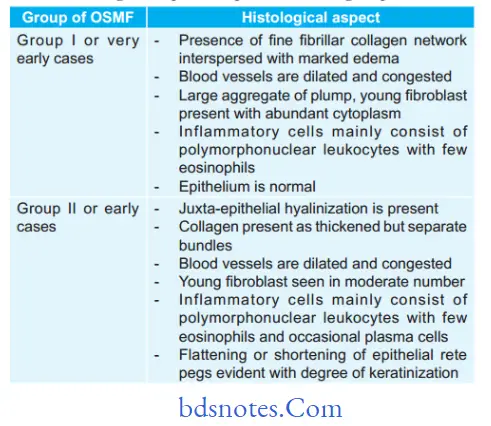
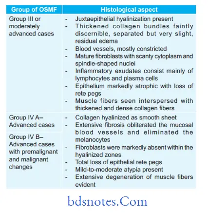
Histopathology Based On Various Types Of OSMF
Khanna JN and Andrade NN in 1995 given the group classification system for OSMF which consists of various groups of OSMF and their clinical and histological aspect. So following are the histological aspects as per the OSMF group:
oral submucous fibrosis microscopic features
“Understanding The Role Of Collagen In Osf Histopathology”
“Comprehensive Overview Of Osf Histopathology And Its Significance”
Leave a Reply