• Skip to main content
  • Skip to secondary menu
  • Skip to primary sidebar
  • About Us
  • Terms of Use
  • Privacy Policy
  • Disclaimer
  • Contact Us
  • Sitemap

BDS Notes

BDS notes, Question and Answers

  • Public Health Dentistry
  • Periodontics
  • Pharmacology
  • Pathology
  • Orthodontics
    • Anchorage In Orthodontics
    • Mandibular Growth, Functional Matrix
    • Retention and Relapse
  • General Surgery
    • Cysts: Types, Causes, Symptoms
    • Maxillofacial Fractures, Disorders, and Treatments
    • Lymphatic Disorders
    • Neurological and Facial Disorders
  • Temporal And Infratemporal Regions
    • Spinal and Neuroanatomy
  • Dental Materials
    • Dental Amalgam
Home » Histopathology of Ewing’s Sarcoma: Cellular and Vascular Features

Histopathology of Ewing’s Sarcoma: Cellular and Vascular Features

July 25, 2025 by Joankessler parkland Leave a Comment

Histopathology of Ewing’s Sarcoma: Cellular and Vascular Features

Question. Write a short note on Ewing’s sarcoma—histopathology.

“Understanding the role of histopathology in diagnosing Ewing’s sarcoma: Q&A explained”

Answer. Following is the histopathology of Ewing’s sarcoma:

Ewing’s sarcoma pathology slides

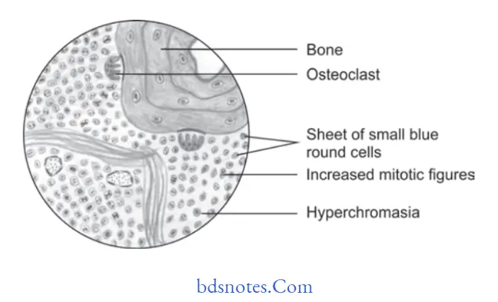
  • The lesion consists of solid sheets of small round cells with very minimal stroma but at a few places, connective tissue septae are present.
  • Cells are round and small in shape along with scanty cytoplasm, nuclei of the cell is large,e round to oval in shape along with dispersed chromatin and hyperchromasia.
  • Borders of cell are ill defied.
  • Cells also show mitotic fiures.
  • Cells of the sarcoma are arranged in fiigree pattrn.
  • Small vascular channels are also evident.
  • Hemorrhage along with vascular lakes or sinuses are appreciated.
  • Perivascular sparing and geographical necrosis is very common in Ewing’s sarcoma.

“Importance of studying histopathology of Ewing’s sarcoma: Questions explained”

Benign and malignant tumors of Oral cavity Ewing’s sarcoma

“Common challenges in identifying Ewing’s sarcoma effectively: FAQs provided”

Filed Under: Oral Pathology

Reader Interactions

Leave a Reply Cancel reply

Your email address will not be published. Required fields are marked *

Primary Sidebar

Recent Posts

  • Branchial Cleft Cyst: Background, Pathophysiology, Etiology
  • Maxillary Nerve: Origin, Course, And Branches
  • The Father Of Anatomy And A Great Anatomist Herophilus
  • Bone Structure – Anatomy
  • The External Carotid Artery: Anatomy, Branches, And Functions
  • Occipitofrontalis Muscle
  • Superficial Temporal Artery
  • Platysma Muscle
  • Cartilage
  • Cauda Equina And Conus Medullaris Syndromes
  • Subcutaneous Injections And Device Management
  • Types Of Circulation: Pulmonary, Systemic, And Portal
  • Structure Of Skeletal Muscle
  • Elastic Cartilage
  • Cellular Organelles And Structure
  • The Golgi Apparatus – The Cell
  • The Cytoplasmic Inclusions Of Certain Plant Cells
  • Dental Abscess
  • Laser Surgery
  • Our Facial Muscles And Their Functions

Copyright © 2026 · Magazine Pro on Genesis Framework · WordPress · Log in