Histological Progression of Dental Caries: Enamel to Dentin Destruction
Question. Defy dental caries. Describe the histopathology of caries in enamel and dentin.
Or
Describe briefly the histopathology of dental caries.
Answer. “Dental caries is an irreversible progressive microbial disease of the calcified tissues of the teeth, characterized by the demineralization of the inorganic portion and distortion of the organic substances of the tooth, which often leads to cavitation”.
“Understanding the role of histology in dental caries progression: Q&A explained”
Histopathological Features Of Caries In Enamel
Early Enamel Caries
- There will be a loss of interprismatic or interrodsubstances with a crease in prominence of these enamel rods.
- Dark lines of often appear right angles to the enamel rods, suggesting segments.
- Accentuation of the incremental striae of Retzus often occurs.
“Importance of studying histological changes in dental caries: Questions explained”
“Common challenges in diagnosing dental caries progression effectively: FAQs provided”
Advanced Enamel Caries
- It presents several zones in the tissues, out of which four zones are visible, starting from the inner advancing front of the lesion, the zones are:
Zone 1: translucent Zone
- It is the deepest zone that lies at the advancing front of the enamel lesion.
- This zone is more porous than normal enamel.
- Pores are larger than normal enamel.
- Pore volume is 1%.
- This zone appears structureless.
- This zone contains more fluoride than normal enamel.
“Steps to explain causes of dental caries progression: Acid production vs demineralization: Q&A guide”
Zone 2: Dark Zone
- The dark zone is located just superficial to the translucent zone,
and its dark appearance is due to the excessive demineralization of the enamel. - The zone is narrower in rapidly advancing caries, and it is wider in slowly advancing lesions.The zone contains 2 to 4% pore volume.
- Pores are larger than normal but smaller than those of the translucent zone.
- This zone reveals some degree of remineralization oof he carious lesion.
Zone 3: body of lesion
- The zone is situated between the dark zone and the surface layer of enamel.
- It represents the area of greatest demineralization.
- Pore volume is 5 to 25%.
- This zone contains appetite crystals larger than those of the normal enamel.
- Large crystals result from the reprecipitation of minerals dissolved from the deeper zone.
“Role of Streptococcus mutans in causing enamel and dentin destruction: Questions answered”
Zone 4: surface Zone
- Surface zone, when examined by the polarizing light, appears relatively
- unaffected; it may be due to the surface remineralization by the salivary mineral ions.
Histological Features Of Caries In Dentin/Dentinal Caries
Dentinal caries histologically presents five zones in the tissue, which are:
Zone 1: normal Dentin
- This zone represents the innermost layer of the carious dentin where the dentinal tubules appear normal.
- There is evidence of fat degeneration of the process.
- No crystals in the lumen of the tubules.
- No bacteria in the tubules.
- Intertubular dentin has normal cross-banded collagen and normal dense apatite crystals.
“Early warning signs of issues addressed by understanding dental caries pathogenesis: Common questions”
Zone 2: sub-transparent Dentin
- This is the zone of dentinal sclerosis and is characterized by the deposition of very fine crystal structures within the dentinal tubules.
- The superficial layer shows areas of demineralization and damage to the odontoblastic processes.
- No bacteria in the tubules.
- Dentin is capable of remineralization.
Zone 3: transparent Dentin
- This zone appears transparent, and this is because of the decalcification of dentin.
- It is softer than normal dentin.
- No bacteria in tubules.
Cross-banded intertubular collagen is still intact. - This zone is capable of self-repair and remineralization.
“Asymptomatic vs symptomatic effects of ignoring dental caries progression: Q&A”
Zone 4: turbid Dentin
- This zone is marked by widening and distortion of dentinal tubules, which are packed with microorganisms.
- There is a very small amount of minerals in dentin, denaturation of collagen fibers also takes place.
- Zone cannot undergo self-repair or remineralization.
- This zone must be removed before restoration.
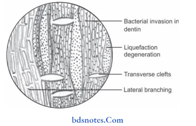
Zone 5: infected Dentin
- This is the outermost zone of the carious dentin.
- It is characterized by complete destruction of dentinal tubules.
- In this zone the area of decomposition of dentin, which occur along the direction of dentinal tubules are called “Liquefaction foci of Miller”, which occur perpendicular to dentinal tubules are called “Transverse clefts”.
- In the process, the entire dentinal structure becomes destroyed, and cavitation begins from the dentino-enamel junction.
Leave a Reply