Histopathological Features Of Shell Teeth
Question. Write a short note on dentinogenesis imperfecta.
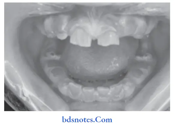
Answer. Dentinogenesis imperfecta is an inherited disorder of dentin formation that affects deciduous and permanent dentition.
Classifiation
“Understanding the role of histopathology in diagnosing shell teeth: Q&A explained”
Sheild’s Classification
- Dentinogenesis imperfecta Type I: Dentinogenesis imperfecta without osteogenesis imperfecta.
- Dentinogenesis imperfecta Type II: Dentinogenesis imperfecta with osteogenesis imperfect
- Dentinogenesis imperfecta Type III: It is a racial isolate in Maryland and is known as Brandywine type.
Extensive studies have shown that dentinogenesis imperfect is a disorder from osteogenesis imperfect, so the following revised classification is given:
- Dentinogenesis imperfecta1: Dentinogenesis imperfect a without osteogenesis imperfecta (opalescent dentine). This corresponds to dentinogenesis imperfect
Type II of Sheild’s classification. - Dentinogenesis imperfect 2: Brandywine type dentinogenesis imperfect: This corresponds to dentinogenesis imperfect Type II of Sheild’s Classification.
“Importance of studying histopathological features of shell teeth: Questions explained”
There is no substitute in present classification for the category which is designated as Dentinogenesis imperfect Type I of Sheild’s classification.
Etiology
Gene affected is present on chromosome 4 and it codes for DSPP (Dentine sialoprotein and phosphoprotein)
“Role of enamel hypoplasia in causing shell teeth: Questions answered”
Clinical Features
- On eruption, the teeth exhibit a normal contour and have an opalescent amber-like appearance.
- A few days after the eruption, the teeth may achieve the normal color. Finally, the teeth become gray or brownish with a bluish reflection from the enamel.
- In some cases, the affected teeth may exhibit hypomineralized areas on the surface of the enamel.
- Teeth are not particularly sensitive even when most of the surface enamel is lost.
- The dentin is soft and easily penetrable in dentinogenesis imperfecta; these teeth are not caries-prone.
“Common challenges in identifying shell teeth effectively: FAQs provided”
Histopathology
- Histopathologically, the enamel appears normal in Dentinogenesis imperfecta. Mantle dentin is also nearly normal.
- Dentinal tubules are fewer in number per square unit area of dentin as compared to normal dentin. The tubules are often distorted, irregular in shape, widely spaced, and larger.
- Pulp chamber and root canal are often obliterated by the abnormal dentin deposition.
- DEJ appears smooth or flattened instead of being scalloped.
- A large area of tubular dentin is present.
“Steps to explain causes of shell teeth: Genetic vs developmental factors: Q&A guide”
Leave a Reply