Histology And Clinical Anatomy Of The Gingiva
Define oral mucosa. Write in detail the microscopic and macroscopic features of the gingiva.
Answer:
Definition:
- The moist lining of the oral cavity that communicates with the exterior is called oral mucosa.
Gingiva:
- It is defined as the tissue that covers the alveolus and encircles the necks of teeth.
- It extends from the dentogingivial junction to the alveolar mucosa.
- It is immovable and firmly attached to the periosteum of the alveolar bone.
Microscopic features:
1. Epithelium:
- Thick stratified squamous epithelium.
- The layers observed are:
- Stratum basale
- Stratum spinosum
- Stratum granulosum
- Stratum corneum.
- It may be ortho-keratinized or para-keratinized.
- It shows stippled surface.
2. Junction between epithelium and lamina propria:
- Convoluted
- Presence of plenty of deep rete pegs that are closely placed.
- This prevents the epithelium from being stripped off.
3. Lamina propria:
- Contains long and narrow connective tissue papillae.
It has a papillary layer and a reticular layer. - Not highly vascular.
- It is made up of collagen bundles, long capillary loops, lymphatics, nerve tissue, and cells like fibroblasts, histocytes, monocytes, mast cells, and lymphocytes.
4. Submucosa:
- A distinct submucosa layer is absent.
- Lamina propria is directly attached to the periosteum.
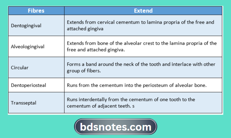
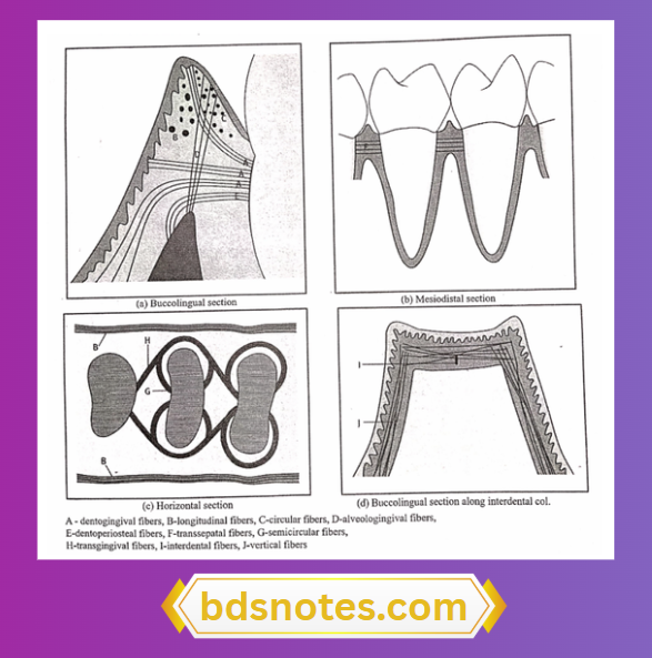
Fibers:
Macroscopic features:
- Colour- coral pink normally but it depends on.
- Thickness of epithelium
- Degree of keratinization
- Degree of pigmentation.
- Amount of circulation.
- Consistency – firm and resilient
- Contour – scalloped
- Surface -stippling surface.
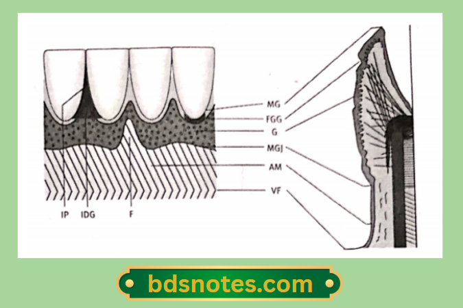
Parts of gingiva:
1. Free or marginal gingiva:
- Embraces the necks of the teeth.
- Width of free gingiva.
- 1 mm – in normal healthy gingiva
Increased in:
- Chronic marginal gingivitis.
- Chronic periodontitis.
- The dividing line between the free gingiva and the attached gingiva is the free gingival groove.
2. Attached gingiva:
- Part of the gingiva that is firmly bound to tire periosteum
- Boundaries.
- Superiorly – free gingival groove.
- Inferiorly – mucogingival line.
- It has a greater width in the maxilla compared to the mandible.
- It is about 4-6 mm.
- Its width decreases in pathological conditions.
- Due to the presence of pockets and gingival recession.
3. Interdental papilla:
- It is that part of the gingiva that fills the space between two adjacent teeth.
- The surface of the interdental papilla is triangular.
- The tip and margins are unattached and the central portion is attached.
Gingival sulcus:
- It is a ‘ V’ shaped space between the marginal gingiva and the tooth surface.
- Its depth is about 0-2 mm normal.
- The base is formed by the superior end of the junctional epithelium.
Leave a Reply