Histological Structure Of Pituitary Gland
“What is the histological structure of the pituitary gland? A detailed question and answers guide”
Histology of Hypophysis Cerebri
Answer:
- It is also called Pituitary gland
- It has been divided into
- Anterior part pars anterior
- Intermediate part pars intermedia
- Posterior partpars posterior
- Pars posterior & infundibular stalk together forms neurohypophysis
- Pars anterior & pars intermedia forms adenohypophysis
“Understanding the histological structure of the pituitary gland through FAQs: Composition, functions, and uses explained”
Histology of hypophysis cerebriv Adenohypophysis:
Histology of hypophysis cerebri Pars anterior:
- It consists of cords of cells separated by Sinusoids
- It consists of following cells
- Chromophil
- Contains brightly stained granules
- It is further classified into
- Chromophil
Histology of hypophysis cerebri Acidophil or Alpha Cells:
Alpha Cells Types:

“Importance of studying the histological structure of the pituitary gland for medical students: Questions explained”
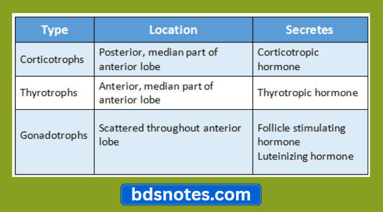
Histology of hypophysis cerebri Basophil or beta cells:
Beta Cells Types:
“Common challenges in mastering the histological structure of the pituitary gland notes effectively: FAQs provided”
2. Chromophobe cells
- Contains very few granules
Pars tuberalis:
- Histology of hypophysis cerebri Pars tuberalis Consists of:
- Undifferentiated cells
- Acidophil
- Basophil
Pars intermedia:
- It is poorly developed
- Contains colloid filled vesicles
- Some cells produce melanocyte stimulating hormone
Pars intermedia Cells:
- Beta cells
- Secretory cells
- Chromophobe cells
“Factors influencing success with pituitary gland studies: Q&A”
Pars intermedia Neurohypophysis:
Pars intermedia Pars posterior:
- It consists of numerous Unmyelinated nerve fibres
- These are axons of neurons
- There are supporting cells called Pituicytes between these axons
- These cells have long dendritic processes
- The pars posterior is associated with the release of
“Steps to explain cell types in the pituitary gland: Acidophils vs basophils vs chromophobes: Q&A guide”
- Vasopressin
- Oxytocin
Leave a Reply