• Skip to main content
  • Skip to secondary menu
  • Skip to primary sidebar
  • About Us
  • Terms of Use
  • Privacy Policy
  • Disclaimer
  • Contact Us
  • Sitemap

BDS Notes

BDS notes, Question and Answers

  • Public Health Dentistry
  • Periodontics
  • Pharmacology
  • Pathology
  • Orthodontics
    • Anchorage In Orthodontics
    • Mandibular Growth, Functional Matrix
    • Retention and Relapse
  • General Surgery
    • Cysts: Types, Causes, Symptoms
    • Maxillofacial Fractures, Disorders, and Treatments
    • Lymphatic Disorders
    • Neurological and Facial Disorders
  • Temporal And Infratemporal Regions
    • Spinal and Neuroanatomy
  • Dental Materials
    • Dental Amalgam
Home » Histological Structure Of Pituitary Gland

Histological Structure Of Pituitary Gland

July 25, 2025 by Kristensmith Taylor Leave a Comment

Histological Structure Of Pituitary Gland

Question 1. Histology of Pituitary Gland
Answer:

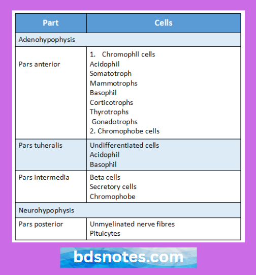
  • It is also called hypophysis cerebri
  • It has been divided into
    • Anterior partpars anterior
    • Intermediate partpars intermedia
    • Posterior partpars posterior
    • Pars posterior & infundibular stalk together forms neurohypophysis
    • Pars anterior & pars intermedia forms adenohypophysis

“Understanding the histological structure of the pituitary gland through FAQs: Composition, functions, and uses explained”

Endocrine Cells Of Pituitary Gland

“Importance of studying the histological structure of the pituitary gland for medical students: Questions explained”

Question 2. Adrenal Cortex Zone Fasciculatum
Answer:

  • It is the middle layer of Suprarenal cortex
  • It forms middle 3/5th of the cortex

Adrenal cortex zone fasciculatum Cells:

  • They are arranged in straight columns
  • Sinusoids intervene between them
  • Cells are large, polyhedral
  • Contains basophilic cytoplasm & vesicular nuclei
  • They are rich in lipids & vitamin C
  • They produce glucocorticoids

“Common challenges in mastering the histological structure of the pituitary gland notes effectively: FAQs provided”

Filed Under: Anatomy

Reader Interactions

Leave a Reply Cancel reply

Your email address will not be published. Required fields are marked *

Primary Sidebar

Recent Posts

  • Branchial Cleft Cyst: Background, Pathophysiology, Etiology
  • Maxillary Nerve: Origin, Course, And Branches
  • The Father Of Anatomy And A Great Anatomist Herophilus
  • Bone Structure – Anatomy
  • The External Carotid Artery: Anatomy, Branches, And Functions
  • Occipitofrontalis Muscle
  • Superficial Temporal Artery
  • Platysma Muscle
  • Cartilage
  • Cauda Equina And Conus Medullaris Syndromes
  • Subcutaneous Injections And Device Management
  • Types Of Circulation: Pulmonary, Systemic, And Portal
  • Structure Of Skeletal Muscle
  • Elastic Cartilage
  • Cellular Organelles And Structure
  • The Golgi Apparatus – The Cell
  • The Cytoplasmic Inclusions Of Certain Plant Cells
  • Dental Abscess
  • Laser Surgery
  • Our Facial Muscles And Their Functions

Copyright © 2026 · Magazine Pro on Genesis Framework · WordPress · Log in