• Skip to main content
  • Skip to secondary menu
  • Skip to primary sidebar
  • About Us
  • Terms of Use
  • Privacy Policy
  • Disclaimer
  • Contact Us
  • Sitemap

BDS Notes

BDS notes, Question and Answers

  • Public Health Dentistry
  • Periodontics
  • Pharmacology
  • Pathology
  • Orthodontics
    • Anchorage In Orthodontics
    • Mandibular Growth, Functional Matrix
    • Retention and Relapse
  • General Surgery
    • Cysts: Types, Causes, Symptoms
    • Maxillofacial Fractures, Disorders, and Treatments
    • Lymphatic Disorders
    • Neurological and Facial Disorders
  • Temporal And Infratemporal Regions
    • Spinal and Neuroanatomy
  • Dental Materials
    • Dental Amalgam
Home » Hairy Leukoplakia: Clinical Features, Histopathology, and Differential Diagnosis

Hairy Leukoplakia: Clinical Features, Histopathology, and Differential Diagnosis

July 23, 2025 by Joankessler parkland Leave a Comment

Hairy Leukoplakia: Clinical Features, Histopathology, and Differential Diagnosis

Question. Write notes on hairy leukoplakia.

Answer. Hairy leukoplakia is HIV associated mucosal disorder,which often involves lateral and ventral surfaces of tongue.

Homosexual man with HIV infection may develop white patchy lesion in oral cavity.

“Understanding the role of hairy leukoplakia in oral health: Q&A explained”

Clinical Features

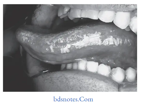
  • Clinically hairy leukoplakia occurs more frequently on the lateral border of the tongue however it can also occur on flor of the mouth, buccal mucosa, etc.
  • The lesion often appears as white patch and is characterized by an irregular surface, exhibiting numerous linear vertical folds or projections, sometimes so marked to as resemble “Hairs”.
  • The lesions are always colonized by Candida albicans.
    Hairy leukoplakia probably occurs as an opportunistic infection caused by Epstein­Barr virus.
  • Hairy leukoplakias are asymptomatic lesions and whenever they occur they occur on buccal mucosa, the lesions are smooth and homogeneous with straitened margin.

“Importance of studying hairy leukoplakia for better diagnostic outcomes: Questions explained”

Histopathology

  • A very characteristic fiding in hairy leukoplakia is presence of subcorneal upper spinous layer zone made up of cytopathologically altered keratinocytes.
  • Parakeratin layer is thick often colonized by candidal hyphae.
  • The submucosa does not exhibit many inflammatory cell infitrate.

Differential Diagnosis

  • Lichen planus
  • Verrucous leukoplakia
  • Chronic tongue biting habits.

“Common challenges in diagnosing hairy leukoplakia effectively: FAQs provided”

Benign and malignant tumors of Oral cavity Hairy leukoplakia

“Role of Epstein-Barr virus in causing hairy leukoplakia: Questions answered”

Filed Under: Oral Pathology

Reader Interactions

Leave a Reply Cancel reply

Your email address will not be published. Required fields are marked *

Primary Sidebar

Recent Posts

  • Branchial Cleft Cyst: Background, Pathophysiology, Etiology
  • Maxillary Nerve: Origin, Course, And Branches
  • The Father Of Anatomy And A Great Anatomist Herophilus
  • Bone Structure – Anatomy
  • The External Carotid Artery: Anatomy, Branches, And Functions
  • Occipitofrontalis Muscle
  • Superficial Temporal Artery
  • Platysma Muscle
  • Cartilage
  • Cauda Equina And Conus Medullaris Syndromes
  • Subcutaneous Injections And Device Management
  • Types Of Circulation: Pulmonary, Systemic, And Portal
  • Structure Of Skeletal Muscle
  • Elastic Cartilage
  • Cellular Organelles And Structure
  • The Golgi Apparatus – The Cell
  • The Cytoplasmic Inclusions Of Certain Plant Cells
  • Dental Abscess
  • Laser Surgery
  • Our Facial Muscles And Their Functions

Copyright © 2026 · Magazine Pro on Genesis Framework · WordPress · Log in