Forearm Upper Limb Anatomy Notes
Front Of Forearm
Question 1. Describe the flexor retinaculum at the wrist in brief.
Answer.
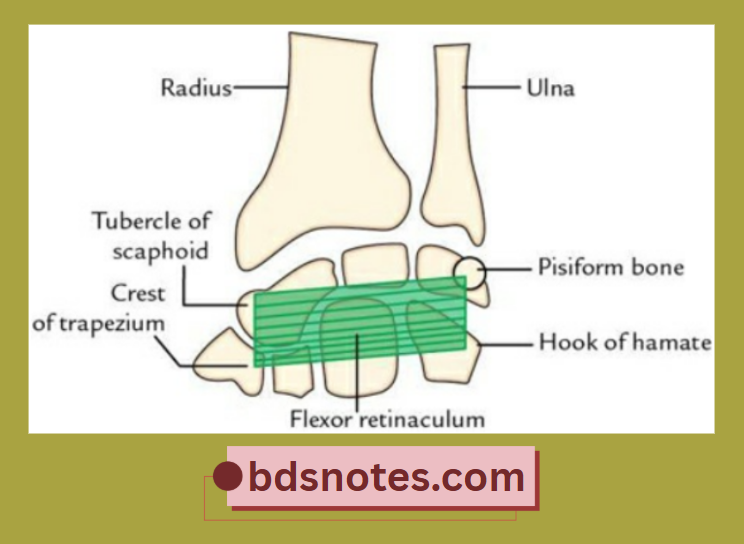
The flexor retinaculum is a strong fibrous band formed by the thickening of deep fascia in front of the carpal bones (anatomical wrist). It bridges the anterior concavity of the carpus and converts it into an osseofibrous tunnel called the carpal tunnel.
Read And Learn More: Selective Anatomy Notes And Question And Answers
Flexor Retinaculum at Wrist Attachments
Flexor Retinaculum at the Wrist is rectangular and attached as follows:
- Medially, to the pisiform bone and the hook of the Hamate
- Laterally, to the tubercle of the scaphoid and the crest of the trapezium
Forearm Upper Limb Anatomy Notes
“Understanding the anatomy of the forearm and upper limb”
“Techniques for managing high-risk groups with injuries”
Flexor Retinaculum at Wrist Features
- Converts the concavity of carpus into an osseofibrous tunnel – the carpal tunnel.
- Proximally, it gives attachment to the tendon of palmaris longus.
- Distally, it gives attachment to the apex of the palmar aponeurosis.
Anatomy of the Forearm Upper Limb
“Global prevalence of forearm injuries in athletes”
Flexor Retinaculum at Wrist Superficial Relations
These are
- Ulnar artery and nerve
- Palmar cutaneous branch of median nerve
- Tendon of palmaris longus
- Superficial palmar branch of radial artery
“Importance of studying forearm anatomy for healthcare professionals”

Leave a Reply