Skeletal Maturity Indicators
“What are Fishman’s Skeletal Maturity Indicators and why do they matter?”
Leonard S Fishman in 1982 had proposed this system.
- Fishman had used four anatomical sites, i.e. location on thumb, third figer, fit figer and radius.
- Eleven discrete adolescent skeletal maturity indicators covering entire period of adolescent development are described.
- Fishman system of interpretation uses four stages of bone maturation which are:
- Epiphysis equal in width to diaphysis
- Appearance of adductor sesamoid of thumb
- Capping of epiphysis
- Fusion of epiphysis.
“Understanding the role of Fishman’s Skeletal Maturity Indicators in orthodontics”
Read And Learn More: Orthodontics Question And Answers
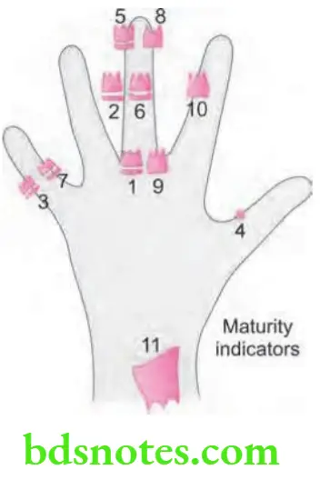
- Following are the Fishman’s skeletal maturity indicators:
- S.M.I 1: The third finger proximal phalanx shows equal width of epiphysis and diaphysis.
- S.M.I 2: Width of epiphysis is equal to that of diaphysis in the middle phalanx of third finger.
- S.M.I 3: Width of epiphysis is equal to that of diaphysis in the middle phalanx of fit finger.
- S.M.I 4: Appearance of adductor sesamoid of the thumb
- S.M.I 5: Capping of epiphysis seen in distal phalanx of third finger.
- S.M.I 6: Capping of epiphysis seen in middle phalanx of third finger.
- S.M.I 7: Capping of epiphysis seen in middle phalanx of fit finger.
- S.M.I 8: Fusion of epiphysis and diaphysis in the distal phalanx of third figer.
- S.M.I 9: Fusion of epiphysis and diaphysis in the proximal phalanx of third figer.
- S.M.I 10: Fusion of epiphysis and diaphysis in the middle phalanx of third figer.
- S.M.I 11: Fusion of epiphysis and diaphysis is seen in the radius.
“Importance of studying Fishman’s Skeletal Maturity Indicators for better outcomes”
S.M.I denotes skeletal maturity indicator.
“Common challenges in applying Fishman’s Skeletal Maturity Indicators effectively”
Leave a Reply