Filtration And Collimation
Write short note on collimation and filtration.
Or
Write short note on filtration and collimation.
Or
Write short note on filtration.
Answer.
Filtration
An X-ray beam has photons of different energies but only the photon with sufficient energy are able to penetrate the anatomic structures and useful in diagnostic radiography. Those photons with low penetrating power (long wavelength) contribute only to patient exposure and not to information on film. Thus for patient safety these photons with low power should be removed. This procedure of increasing the quality of radiation by removal of low power photon is known as “Filtration”.
It is done by placing an aluminum filter in path of beam.
“Understanding the role of filtration and collimation in X-ray imaging: Q&A explained”
Type of Filtration
- Inherent filtration.
- Added filtration
- Total filtration
Inherent Filtration
Filtration provided by materials of X-ray tube head, e.g. glass, insulating oil and the tube head seal. It is equivalent to 0.5 to 1.5 mm of aluminum.
“Common challenges in applying filtration and collimation effectively: FAQs provided”
Added Filtration
As the name suggests additional, an additional aluminum disk is placed between the tube head seal and collimator in the path of primary beam. It is 0.5 mm thick.
Total Filtration
- Total filtration is the sum of Inherent filtration and Added filtration.
- At or below 70 kVp requires minimum total filtration of l.5 mm of aluminum thickness.
- Above 70 kVp require a minimum total filtration of 2.5 mm of aluminum thickness.
- Contrast and quality of films get increase with the use of filter.
“Importance of studying filtration and collimation for better radiation safety: Questions explained”

“Steps to explain different types of filtration: Inherent vs added filtration: Q&A guide”
Collimation
Collimation is the process of restriction the size of the X-ray beam and thus the volume of irradiated tissue of the patient from which the scattered photons originate.
- It allows only a useful beam to emerge (the useful beam is defined as that part of the primary radiation which is allowed to emerge through the collimating device).
“Role of aluminum filtration in reducing soft radiation: Questions answered”
- When an X-ray beam falls on tissue 90% of it is absorbed by tissue and 10% result in scattered beam and travels in all direction.
- The scattered radiation does not contribute to information but only adds to film fog and degrades the image.
- The collimation decreases the risk of radiation, minimizes scattered radiation and decreases the fog with sharper image and better contrast.
- In intraoral machines, there are fixed collimators and in extraoral machines, they are adjustable.
Uses of collimation
- It reduces the volume of irradiated tissues and decreases the radiation exposure to patient.
- It reduces the size of X-ray beam and amount of scattered photons.
- It reduces the film fog and enhances the quality of image.
Types of collimators
Following are the type of collimators:
“Early warning signs of issues caused by improper filtration and collimation: Common questions”
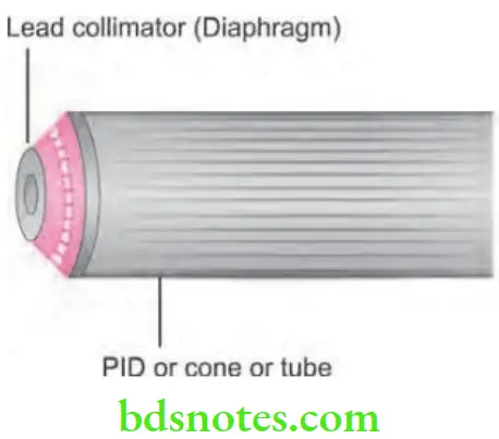
Diaphragm Collimator
- It consists of a thick plate of radiopaque material with opening in it.
- Collimator should lie over the port in X-ray head by which X-ray beam emerges.
- Aperture should be of different size and shape depending on requirement.
Tubular Collimator
- It is a tube which is manufactured by radiopaque material.
- Combination of diaphragm type and tubular type is used.
- The combination helps in reducing the penumbra at periphery of image.
- Longer the tube small is penumbra.
“Asymptomatic vs symptomatic effects of ignoring proper filtration and collimation protocols: Q&A”
Rectangular Collimator
- They limit the size and they are larger than size of an X-ray film.
- It can be incorporated in a film-holding device.
Slit Collimator
- It is used in OPG machines.
- In CT machines, collimators are of two types i.e.
- Source collimator: It lies in front of the X-ray tube and reduces the beam of radiation to form maximum required fan beam and determines the emitted dose.
- Detector collimator: It is positioned directly in front of the detector and is used to shield detector against scattered radiation and prevent artifacts.
Leave a Reply