Fibroma of the Oral Cavity: Clinical Features, Histology, and Management
Question. Classify the nonodontogenic tumors of the oral cavity and describe fibroma.
Or
Write a short answer on Forma.
Answer.
Fibroma
Fibroma is a benign tumor of connective tissue origin.
“Understanding the role of fibroma in oral health: Q&A explained”
Fibroma Clinical Features
- It is a slow growing lesion and can be seen during 3rd, 4th and 5th decades of life.
- Female predilection is seen.
- It can occur anywhere in the oral cavity but most commonly it is seen on buccal mucosa along plane of occlusion. It also affcts gingiva, tongue, buccal mucosa,lips and palate.
- Lesion appears as elevated nodule of normal color with smooth surface and a sessile or at times pedunculated base.
- Its size can ranges from several millimeters to centimeters.
- Lesion if traumatized it become painful.
- Color of the lesion is pink and texture is smooth.
- Consistency of lesion can be soft or fim or can be elastic.
- At times lesion is traumatized and become inflmed and show ulceration or hyperkeratosis.
“Importance of studying fibroma for better diagnostic outcomes: Questions explained”
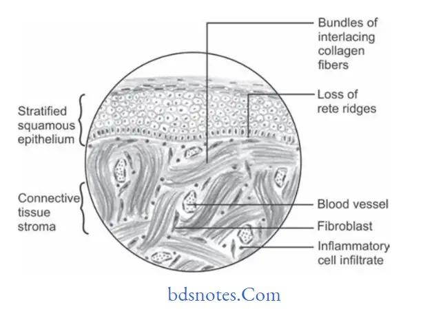
Fibroma Histopathology
- Lesion consists of stratifid squamous epithelium which show shortening and fitting of rete pegs.
- Underlying connective tissue stroma show bundles of interlacing collagen fiers which are interspersed with numerous firoblasts.
- There is presence of chronic inflammatory cell infitrate
consisting of lymphocytes and plasma cells. - Areas of calcification and ossifiation can also be seen.
“Common challenges in diagnosing fibroma of the oral cavity effectively: FAQs provided”
“Differential applications of medical vs surgical treatments: Q&A”
Fibroma Treatment
Excision of the lesion should be done.
Leave a Reply