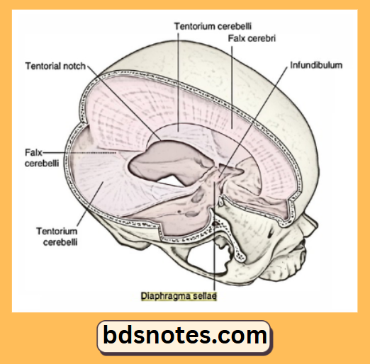
• Skip to main content
  • Skip to secondary menu
  • Skip to primary sidebar
  • About Us
  • Terms of Use
  • Privacy Policy
  • Disclaimer
  • Contact Us
  • Sitemap

BDS Notes

BDS notes, Question and Answers

  • Public Health Dentistry
  • Periodontics
  • Pharmacology
  • Pathology
  • Orthodontics
    • Anchorage In Orthodontics
    • Mandibular Growth, Functional Matrix
    • Retention and Relapse
  • General Surgery
    • Cysts: Types, Causes, Symptoms
    • Maxillofacial Fractures, Disorders, and Treatments
    • Lymphatic Disorders
    • Neurological and Facial Disorders
  • Temporal And Infratemporal Regions
    • Spinal and Neuroanatomy
  • Dental Materials
    • Dental Amalgam
Home » Falx Cerebri

Falx Cerebri

July 26, 2025 by Kristensmith Taylor Leave a Comment

Falx Cerebri

Question 1. Falx cerebri
Answer:

  • It is large sickle-shapes fold of duramater
  • It occupies the median longitudinal fissure between the two cerebral hemispheres

“Understanding falx cerebri through FAQs: Anatomy, functions, and uses explained”

Ends:

1. Anterior end

  • It is narrow & attached to the crista galli

2. Posterior end

  • It is broad & attached to the upper surface of the tentorium cerebelli

Falx cerebri anatomy

Margins:

1. Upper convex margin

  • Attached to the lips of the sagittal sulcus

“Importance of studying falx cerebri for medical students: Questions explained”

2. Lower concave margin

  • It is free

Surfaces:

  • Left & right related to the medial surface of the cerebral hemisphere

Location of falx cerebri

Venous sinuses present in it:

  • Superior Sagittal sinus
  • Inferior Sagittal sinus
  • Straight sinus

“Common challenges in mastering falx cerebri notes effectively: FAQs provided”

Falx Cerebri Tentorium Cerebelli Diagram

“Factors influencing success with falx cerebri studies: Q&A”

Question 2. Meckel’s cave
Answer:

  • It is a recess of duramater present i.r.t the attached margin of the tentorium

Formed by:

  • Evagination of the inferior layer of the tentorium over the trigeminal impression on the petrous temporal bone

Content:

  • Trigeminal ganglion

“Steps to explain the anatomy of the falx cerebri: Dural attachment vs meningeal layers: Q&A guide”

Question 12. Emissary vein
Answer:

  • These veins connect intracranial venous sinuses with extracranial veins
  • They try to relieve raised intracranial pressure

“Role of the falx cerebri in dividing cerebral hemispheres: Questions answered”

Applied anatomy:

  • Infection may reach through the emissary veins into cranial venous sinuses

Various emissary veins:

  • Parietal emissary vein Mastoid emissary vein Condylar emissary vein

Filed Under: Anatomy

Reader Interactions

Leave a Reply Cancel reply

Your email address will not be published. Required fields are marked *

Primary Sidebar

Recent Posts

  • Branchial Cleft Cyst: Background, Pathophysiology, Etiology
  • Maxillary Nerve: Origin, Course, And Branches
  • The Father Of Anatomy And A Great Anatomist Herophilus
  • Bone Structure – Anatomy
  • The External Carotid Artery: Anatomy, Branches, And Functions
  • Occipitofrontalis Muscle
  • Superficial Temporal Artery
  • Platysma Muscle
  • Cartilage
  • Cauda Equina And Conus Medullaris Syndromes
  • Subcutaneous Injections And Device Management
  • Types Of Circulation: Pulmonary, Systemic, And Portal
  • Structure Of Skeletal Muscle
  • Elastic Cartilage
  • Cellular Organelles And Structure
  • The Golgi Apparatus – The Cell
  • The Cytoplasmic Inclusions Of Certain Plant Cells
  • Dental Abscess
  • Laser Surgery
  • Our Facial Muscles And Their Functions

Copyright © 2026 · Magazine Pro on Genesis Framework · WordPress · Log in