Exploring The Digastric Triangle: Anatomy, Function, And Clinical Significance
Question 1. Mention the branches of lingual artery
Answer:

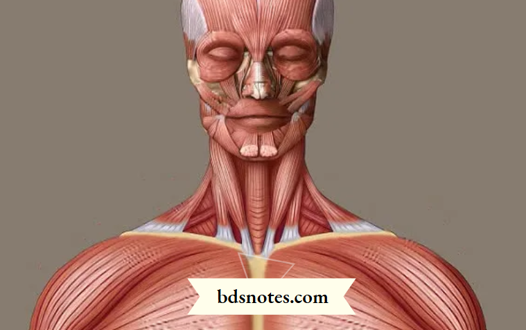
The digastric triangle, also known as the submandibular triangle, is a significant anatomical region in the neck. It is bounded by the muscles of the digastric muscle and the mandible, and it plays a vital role in various functions such as swallowing and speaking. In this article, we will explore the anatomy, function, innervation, blood supply, clinical significance, relations, and variations of the digastric triangle. Understanding this area is crucial for both medical professionals and students alike.
Digastric Triangle Key Significance
- The digastric triangle is bordered by the digastric muscle and the mandible, forming a key area in the neck.
- It contains important structures such as the submandibular gland, lymph nodes, and major blood vessels.
- The digastric muscle helps in depressing the mandible and elevating the hyoid bone during swallowing.
- Innervation of the digastric triangle comes from the mylohyoid nerve and the digastric branch of the facial nerve.
- Variations in the digastric triangle can impact surgical procedures and anatomical studies.
Anatomy Of The Digastric Triangle
Borders Of The Triangle
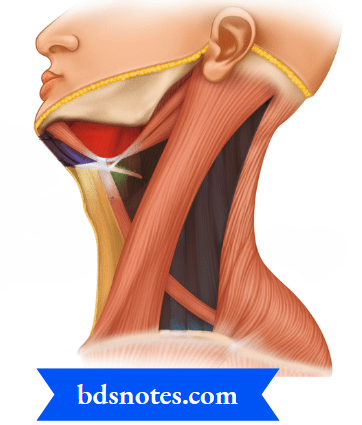
Okay, so the digastric triangle, also known as the submandibular triangle, is this little area in your neck that’s actually pretty important. It’s defined by a few key boundaries. Think of it like a little anatomical neighborhood. The anterior belly of the digastric muscle makes up one side, and the posterior belly of the digastric and the stylohyoid muscles form another. The inferior border of the mandible (your jawbone) makes up the top side. It’s like a muscular little pocket there.
Muscles Involved
Obviously, the main muscle here is the digastric. It’s called that because “di” means two and “gaster” means belly, so it literally has two bellies. The digastric muscle divides the anterior triangle of the neck into smaller divisions like the carotid triangle, the submental triangle and the submandibular triangle. But there are other muscles that play a role, too. The mylohyoid and hyoglossus muscles form the floor of the triangle. These muscles help with things like swallowing and speaking. The stylohyoid muscle runs along the posterior belly of the digastric and assists in elevating the hyoid bone.
Floor And Roof Structures
So, what’s underneath and on top of this triangle? The floor is made up of the mylohyoid and hyoglossus muscles, like I mentioned. These muscles are important for supporting the tongue and helping with swallowing. The roof is formed by the skin, the platysma muscle (that thin muscle in your neck), and some layers of fascia (connective tissue). Inside, you’ll find the submandibular gland and lymph nodes, which are part of your immune system. It’s a pretty packed space!
Think of the digastric triangle as a 3D space. It’s not just about the borders, but also what’s deep inside and what covers it. This is important for surgeons and doctors to know, so they don’t accidentally damage anything important during procedures.
Function Of The Digastric Triangle
Mandible Depression
The digastric muscle, a key component of the digastric triangle, plays a role in depressing the mandible, or opening the mouth. This action is particularly important when resistance is encountered, such as during chewing. The anterior belly pulls the mandible down and back, while the posterior belly assists in stabilizing the hyoid bone, allowing for controlled movement. It’s not the strongest muscle for this action, but it’s a helper, especially when you need to open your mouth wide.
Hyoid Elevation
Another important function is the elevation of the hyoid bone. This is crucial for swallowing and speech. When the mandible is fixed, the digastric muscle can elevate the hyoid bone and larynx. This action shortens the floor of the mouth and increases pressure within the oral cavity, which is essential for propelling food bolus into the pharynx. The digastric muscle works with other suprahyoid muscles to achieve this.
Role In Swallowing
The digastric triangle and its contents are integral to the swallowing process. The coordinated action of the digastric muscle, along with other suprahyoid and infrahyoid muscles, ensures the safe and efficient passage of food from the mouth to the esophagus. Here’s a breakdown:
- Oral Phase: The digastric muscle helps stabilize the hyoid bone, allowing the tongue to propel the bolus backward.
- Pharyngeal Phase: Elevation of the hyoid bone and larynx by the digastric muscle helps to close the airway and open the esophagus.
- Esophageal Phase: While the digastric muscle doesn’t directly participate in this phase, its earlier actions have set the stage for successful swallowing.
The digastric muscle’s role in swallowing is more about setting the stage for the main event. It helps position things correctly so that other muscles can do their jobs effectively. Without it, swallowing could become difficult or even dangerous, increasing the risk of choking.
Think of it like this: the digastric muscle is part of the team that makes sure food goes down the right pipe!
Innervation Of The Digastric Triangle
Anterior Belly Innervation
The anterior belly of the digastric muscle gets its nerve supply from the nerve to the mylohyoid. This nerve is a branch of the inferior alveolar nerve, which itself comes from the mandibular nerve. So, it’s a bit of a chain reaction to get the signal there!
Posterior Belly Innervation
Now, the posterior belly is a different story. It’s innervated by the digastric branch of the facial nerve (cranial nerve VII). This difference in innervation is because the two bellies develop from different pharyngeal arches during development. Pretty neat, huh?
Nerve Pathways
Understanding the nerve pathways is key for diagnosing certain conditions. The facial nerve, after exiting the skull, sends a branch specifically to the posterior digastric belly. The mylohyoid nerve, branching off the inferior alveolar, directly stimulates the anterior belly. Damage to either of these nerves can affect the function of the digastric muscle, impacting things like swallowing and speech.
It’s important to remember that the digastric muscle plays a role in both depressing the mandible and elevating the hyoid bone. Therefore, any nerve damage can lead to noticeable functional deficits. Knowing which nerve innervates which belly helps pinpoint the location of the injury.
Blood Supply To The Digastric Triangle
Arterial Supply
The digastric triangle gets its blood from a few different arteries, which is pretty important for keeping everything in that area working right. The facial artery is a big player here, winding its way through the triangle to supply blood to the submandibular gland and nearby muscles. You’ll also find the submental artery, a branch of the facial artery, contributing to the blood flow in the region. The lingual artery also pitches in, ensuring that the tongue and surrounding structures get the oxygen and nutrients they need. Understanding these arterial pathways is key for surgeons and anyone dealing with neck anatomy.
Venous Drainage
Just like arteries bring blood in, veins take it away. The venous drainage of the digastric triangle primarily involves the facial vein and its tributaries. The facial vein receives blood from smaller veins like the submental and lingual veins, mirroring the arterial supply. These veins eventually drain into the internal jugular vein, a major vessel in the neck. Proper venous drainage is essential to prevent congestion and maintain healthy tissue function. It’s all connected, you know?
Significance Of Blood Vessels
The blood vessels within the digastric triangle aren’t just there for show; they’re super important for a bunch of reasons. First off, they keep the muscles, glands, and other tissues in the area alive and kicking. Second, they can be important landmarks during surgery. Surgeons need to know where these vessels are to avoid damaging them. Plus, problems with these blood vessels, like blockages or aneurysms, can cause serious health issues. So, yeah, these vessels are kind of a big deal. The anterior triangle of the neck contains major arteries originating from the common carotid arteries.
Think of the digastric triangle as a busy intersection for blood vessels. Knowing the routes and connections is vital for anyone working in or studying this area of the neck. It’s not just about memorizing names; it’s about understanding how everything works together to keep us healthy.
Clinical Significance Of The Digastric Triangle
The digastric triangle, while small, is super important because of all the stuff packed inside. Knowing its anatomy is key for surgeons and doctors dealing with neck issues. It’s like a mini-hub for important structures, so problems here can have big effects.
Surgical Considerations
When surgeons work in the neck, the digastric triangle is often a landmark. It helps them find and protect important nerves and blood vessels. For example, during a neck dissection (removing lymph nodes, often for cancer treatment), surgeons need to be super careful around the internal jugular vein and carotid artery, both of which are close by. Understanding the triangle’s boundaries helps avoid accidental damage. Also, procedures involving the submandibular gland often require careful dissection within this area.
Pathologies Associated
Lots of different problems can show up in the digastric triangle. These include:
- Infections: The submandibular lymph nodes, located in this triangle, can get infected and swollen, like with the submental triangle. This is common with things like strep throat.
- Tumors: Both benign and cancerous tumors can occur in the triangle, affecting the salivary glands or lymph nodes. Accurate diagnosis is key.
- Cysts: Branchial cleft cysts, which are birth defects, can sometimes show up in this area. They usually present as a painless lump.
Problems in the digastric triangle can sometimes be tricky to diagnose because the symptoms can overlap with other conditions. A thorough exam and imaging are often needed to figure out what’s going on.
Diagnostic Importance
Feeling around (palpating) the digastric triangle is a standard part of a neck exam. Doctors do this to check for:
- Swollen lymph nodes: This can point to infection, inflammation, or even cancer.
- Masses: Any unusual lumps need to be checked out to rule out tumors or cysts.
- Tenderness: Pain in the area can suggest infection or inflammation.
Imaging tests, like ultrasound or CT scans, can give a better look at the structures inside the triangle. These are helpful for diagnosing problems that can’t be felt during a physical exam. For example, an ultrasound can help distinguish between a cyst and a solid tumor. Also, knowing the anterior belly innervation is important for diagnosing nerve-related issues in the area.
Relations Of The Digastric Triangle
Adjacent Structures
The digastric triangle, also known as the submandibular triangle, doesn’t exist in isolation. It’s snuggled right next to other important areas in the neck. Think of it as a neighborhood, with each area having its own role but also influencing the others. The anterior triangle of the neck is divided by the digastric muscle into the carotid, submental, and submandibular triangle. The stylohyoid muscle also plays a role in defining the borders superiorly.
Neurovascular Relationships
This triangle is a hotspot for nerves and blood vessels. It’s like a major intersection where important routes converge. The facial artery and vein, submental artery and vein, lingual arteries and veins, mylohyoid nerve, and the hypoglossal nerve (CN XII) all hang out here. The digastric muscle itself is closely related to the internal jugular vein, external and internal carotid arteries, and vagus, glossopharyngeal, and hypoglossal nerves. These structures usually pass deep to the posterior belly of the digastric muscle.
Clinical Implications
Understanding the relationships of the digastric triangle is super important for doctors, especially surgeons. Because so many important structures are packed into a small space, any procedure in this area needs to be done with care. For example, surgeons need to be aware of the location of the facial artery when removing a submandibular gland. Also, the triangle’s proximity to major nerves means that nerve damage is a risk during surgery.
Basically, if you’re poking around in the digastric triangle, you need to know exactly what’s next to what. Otherwise, you could end up causing some serious problems. It’s like trying to fix a watch with a hammer – not a good idea.
Here’s a quick rundown of key relationships:
- Carotid Triangle: The posterior belly of the digastric muscle forms the superior border.
- Submental Triangle: Bordered laterally by the anterior bellies of the digastric muscles.
- Submandibular Triangle: Contains the submandibular gland and lymph nodes.
Variations In The Digastric Triangle
Anatomical Variations
Okay, so the digastric triangle isn’t always the same size or shape in everyone. You might find differences in the position of the digastric muscle bellies, which can alter the triangle’s borders. Sometimes, the stylohyoid muscle inserts differently, affecting the posterior border. These variations are usually no big deal, but surgeons need to know about them.
Functional Variations
How the digastric muscle works can also vary. Some people might have a stronger or weaker contraction of one belly compared to the other. This can affect how they depress their mandible or elevate their hyoid bone. These functional differences are often subtle and might not even be noticeable, but they’re there. For example, someone might have a slightly different swallowing pattern because of it.
Knowing about these variations is super important for surgery in the neck area. If a surgeon isn’t aware that someone’s neck anatomy is a bit different, they could accidentally damage something important. For example, the location of the facial artery or the hypoglossal nerve might be slightly different than expected. So, pre-operative imaging and careful dissection are key to avoiding problems. It’s all about being prepared for anything!
Wrapping Up The Digastric Triangle
In summary, the digastric triangle is more than just a part of neck anatomy; it plays a key role in various functions like swallowing and speaking. Understanding its structure helps us appreciate how the muscles and nerves work together in this area. Plus, knowing about the triangle’s clinical significance can aid in diagnosing and treating conditions that affect the neck. So, whether you’re a student, a healthcare professional, or just curious about human anatomy, the digastric triangle is definitely worth a closer look.
Digastric Triangle Frequently Asked Questions
Question 1. What Is The Digastric Triangle?
Answer: The digastric triangle, also known as the submandibular triangle, is an area in the neck formed by muscles and bones. It contains important glands, nerves, and blood vessels.
Question 2. What Are The Main Muscles In The Digastric Triangle?
Answer: The main muscles are the anterior and posterior bellies of the digastric muscle, and the stylohyoid muscle. These muscles help with movements like chewing.
Question 3. How Does The Digastric Triangle Help With Swallowing?
Answer: The digastric triangle plays a role in swallowing by helping to lift the hyoid bone, which is important for moving food down the throat.
Question 4. What Nerves Are Involved In The Digastric Triangle?
Answer: The anterior belly of the digastric muscle is innervated by the mylohyoid nerve, while the posterior belly gets its nerve supply from the facial nerve.
Question 5. What Blood Vessels Are Found In The Digastric Triangle?
Answer: The triangle contains the facial artery and the occipital artery, which supply blood to the muscles and glands located in this area.
Question 6. Why Is The Digastric Triangle Important In Surgery?
Answer: Surgeons need to know about the digastric triangle because it contains important structures. Understanding its anatomy helps prevent damage during operations in the neck.
Leave a Reply