Essential Diagnostic AIDS In Orthodontics
Essential Diagnostic Aids
They are clinical aids that are considered very important for all cases.
- They are simple and do not require expensive equipments.
- Following are the essential diagnostic aids:
- Case history
- Clinical examination
- Study models
- Certain radiographs (lOPA, Bitewing, Panoramic)
- Facial photographs.
“Understanding the role of diagnostic aids in orthodontic treatment planning”
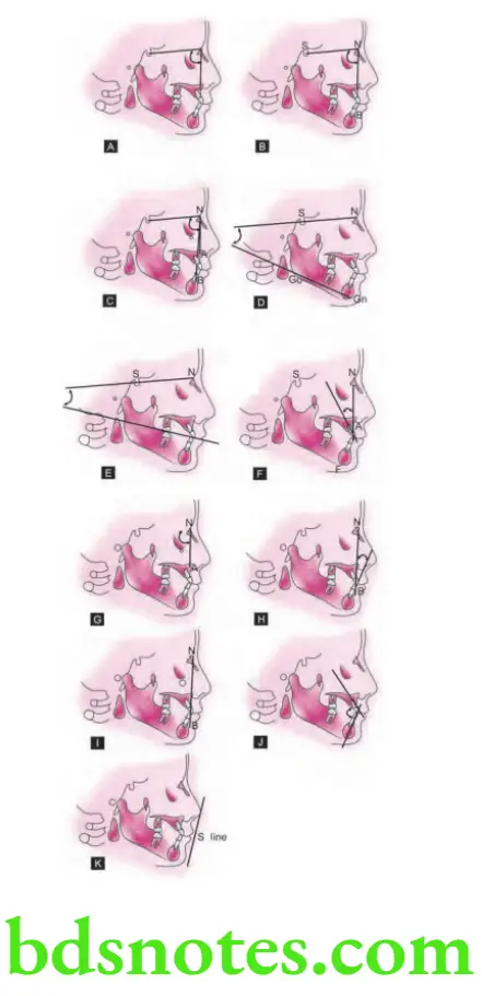
Steiner Analysis
It is a cephalometric analysis.
Cecil C Steiner in the year 1930 developed this analysis.
The Steiner analysis is divided into three parts:
- Skeletal analysis.
- Dental analysis.
- Soft tissue analysis.
“Importance of studying diagnostic aids for better orthodontic success”
Skeletal Analysis
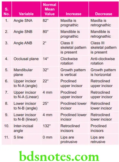
- SNA angle: The angle formed by the intersection of SN plane and a line joining nasion and point A. Indicates position of maxilla in relation to cranium.The mean value is 82°.Value increased in prognathic maxilla (Class 2). Value decreased in retrognathic maxilla (Class 3).
- SNB angle: Angle between SN plane and line joining nasion to point B.
- This angle indicates the position of mandible to cranial base.
- Average value is 80°.
- Value increase in prognathic mandible (Class 3).
- Value decreased in retrognathic mandible (Class 2).
- ANB angle: Angle between line joining point A to nasion and a line joining point B to nasion.
- It indicates position of maxilla and mandible to each other.
- Average value is 2 degree.
- Increased value indicates class II skeletal malocclusion.
- Decreased value indicates class III skeletal malocclusion.
“Common challenges in using essential diagnostic aids effectively”
“Steps to explain different types of essential diagnostic aids in orthodontics”
- Mandibular plane angle: It is the angle between SN plane and mandibular plane.
- Average value is 30°.
- Indicates growth pattrn of individual.
- Lower angle indicates horizontal growing pattrn of individual.
- Increased angle indicates vertical growing pattrn of individual.
- Occlusal plane angle: The angle between the SN plane and occlusal plane.
- Occlusal plane is line passing through the overlapping cusps of the fist premolar and fist molar.
- It has a mean value of 14.5°.
- It indicates the relation of occlusal plane to the cranium and face.
- Also indicates growth pattrn in individual.
Dental Analysis
Maxillary Incisor Position
- Upper incisor is related to N-A line for determination of its position.
- Upper incisor to N-A (Angle):It is the angle formed by the intersection of long axis of the upper central incisor and the line joining nasion to point A. Mean value is 22°.
- An increase in angle indicates proclined upper incisors (class II malocclusion) and decrease in the angle is suggestive of retroclination.
- Upper incisors to N-A (Linear): It is a linear measurement between the labial surface of upper central incisor and the line joining nasion to point A. Mean value is 4 mm.
- It increases with proclined upper central incisor and decreases with retroclination.
“Asymptomatic vs symptomatic effects of misdiagnosed orthodontic cases”
Mandibular Incisor Position
- Lower incisor is related to N-A line for determination of its position.
- Lower Incisor to N-B (angle): The angle between the N-B plane and the long axis of the lower incisor. Mean value is 25°.
- Increased value indicates proclination of lower incisor while the decreased value indicates upright or retroclined lower incisor.
- Lower Incisor to NB (linear): Linear distance between the labial surface of lower central incisor and the line joining nasion to point B. Mean value is 4 mm.
- Increased value indicate proclined lower central incisor while decreased value indicates of retroclination.
“Role of radiographic imaging in orthodontic diagnostics”
Interincisal Angle
- Angle formed between the long axis of the upper and lower central incisor.
- Mean value is 132°
- When upper and lower incisors are proclined angle is acute.
- When upper and lower incisors are retroclined angle is obtuse.
- These angulations help in detecting incisors with defective angulations.
Soft Tissue Analysis
- In well balanced faces, lip lie along the S line.
- Lips which are located anterior to S line are protrusive. Orthodontic treatment should be carried in order to reduce protrusion.
“Early warning signs of untreated issues detectable via diagnostic aids”
Summary of Steiner’s Analysis
Leave a Reply