• Skip to main content
  • Skip to secondary menu
  • Skip to primary sidebar
  • About Us
  • Terms of Use
  • Privacy Policy
  • Disclaimer
  • Contact Us
  • Sitemap

BDS Notes

BDS notes, Question and Answers

  • Public Health Dentistry
  • Periodontics
  • Pharmacology
  • Pathology
  • Orthodontics
    • Anchorage In Orthodontics
    • Mandibular Growth, Functional Matrix
    • Retention and Relapse
  • General Surgery
    • Cysts: Types, Causes, Symptoms
    • Maxillofacial Fractures, Disorders, and Treatments
    • Lymphatic Disorders
    • Neurological and Facial Disorders
  • Temporal And Infratemporal Regions
    • Spinal and Neuroanatomy
  • Dental Materials
    • Dental Amalgam
Home » Esophagus

Esophagus

July 25, 2025 by Kristensmith Taylor Leave a Comment

Esophagus

Histology of Esophagus
Answer:

  • Oesophagus is a tube
  • Histology of esophagus It has four layers

“Factors influencing success with esophagus studies: Q&A”

    1. Mucosa
      • It shows several longitudinal folds that disappear when the tube is distended It is lined by stratified Squamous epithelium
      • Finger like processes project from connective tissue into the epithelium
      • This prevents separation of epithelium
      • Lamina propria contains some mucous glands
      • The muscularis mucosae is absent or poorly developed
    2. Submucosa
      • It contains compound tubuloalveolar mucous glands
      • These glands are present at the level of bifurcation of the trachea
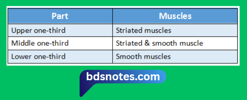
    3. Muscle layer
      • It consists of usual circular & longitudinal layers
    4. External adventitia
      • It is dense fibrous tissue surrounding the muscle layer

“Understanding the esophagus through FAQs: Anatomy, functions, and uses explained”

Esophagus Muscle Layers Histology

“Importance of studying the esophagus for medical students: Questions explained”

Filed Under: Anatomy

Reader Interactions

Leave a Reply Cancel reply

Your email address will not be published. Required fields are marked *

Primary Sidebar

Recent Posts

  • Branchial Cleft Cyst: Background, Pathophysiology, Etiology
  • Maxillary Nerve: Origin, Course, And Branches
  • The Father Of Anatomy And A Great Anatomist Herophilus
  • Bone Structure – Anatomy
  • The External Carotid Artery: Anatomy, Branches, And Functions
  • Occipitofrontalis Muscle
  • Superficial Temporal Artery
  • Platysma Muscle
  • Cartilage
  • Cauda Equina And Conus Medullaris Syndromes
  • Subcutaneous Injections And Device Management
  • Types Of Circulation: Pulmonary, Systemic, And Portal
  • Hierarchical Organization Of Skeletal Muscle Tissue
  • Elastic Cartilage Histology Short Note For Medical Exams
  • Cellular Organelles And Structure
  • The Golgi Apparatus – The Cell
  • The Cytoplasmic Inclusions Of Certain Plant Cells
  • Dental Abscess
  • Laser Surgery
  • Our Facial Muscles And Their Functions

Copyright © 2026 · Magazine Pro on Genesis Framework · WordPress · Log in