Epithelial Attachment: The Tooth-Gum Connection
Question 1. Write briefly about epithelial attachment.
Answer:
Epithelial attachment:
- It is shown by Stem and confirmed by Listgarten and Schroeder,
- They showed the mode of attachment of the ameloblasts to the tooth, to be basal lamina to which hemidesmosomes are attached.
- This is referred to as epithelial attachment.
- Both reduced ameloblasts and gingival epithelial cells form basal lamina on enamel and cementum
- Hemidesmosomes of these cells attach to the basal lamina.
- This basal lamina is referred to as the internal basal lamina.
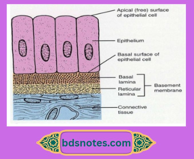
- The lamina propria below the junctional epithelium keeps the epithelial cells of the junctional epithelium immature so that it can develop hemidesmosomes and attach to the tooth.
- They then migrate over it, with their attachment being maintained by the hemidesmosomes.
- The hemidesmosomes hold the cells to the basal lamina so that the strength of the attachment is not diminished despite the migration.
Question 2. Circumvallate papillae.
Answer:
- Location: Just anterior to the sulcus terininalis.
- Number: 8 – 12 in number.
Structure:
- Large round structures.
- They do not protrude above the surface
- They are surrounded by a deep, circular groove for the opening of ducts of minor salivary glands.
- They contain a connective tissue core covered by a keratinized epithelium.
Surfaces;
- Free surface – shows numerous secondary papillae covered by a thin, smooth epithelium.
- Lateral surface – contains numerous taste buds.
Functions:
- Wash out the soluble elements of food
- The main source of salivary lipase.
Leave a Reply