• Skip to main content
  • Skip to secondary menu
  • Skip to primary sidebar
  • About Us
  • Terms of Use
  • Privacy Policy
  • Disclaimer
  • Contact Us
  • Sitemap

BDS Notes

BDS notes, Question and Answers

  • Public Health Dentistry
  • Periodontics
  • Pharmacology
  • Pathology
  • Orthodontics
    • Anchorage In Orthodontics
    • Mandibular Growth, Functional Matrix
    • Retention and Relapse
  • General Surgery
    • Cysts: Types, Causes, Symptoms
    • Maxillofacial Fractures, Disorders, and Treatments
    • Lymphatic Disorders
    • Neurological and Facial Disorders
  • Temporal And Infratemporal Regions
    • Spinal and Neuroanatomy
  • Dental Materials
    • Dental Amalgam
Home » Endochondral Ossification: Step-By-Step Microscopic Changes

Endochondral Ossification: Step-By-Step Microscopic Changes

February 6, 2026 by Kristensmith Taylor Leave a Comment

Endochondral Ossification: Step-By-Step Microscopic Changes

Question 1. Microscopic picture of endochondrial ossification
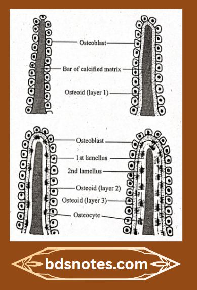
Answer:

  • Following changes occurs during endochondrial ossification
  • Mesenchymal cells are closely packed to form Mesenchymal condensation
  • Some of them differentiate into chondrocytes & lay down hyaline cartilage
  • Cells of this cartilage are
    1. Initially-small & irregular
    2. Later-Enlarges
      • Next, due to lack of nutrition, cells die forming empty spaces called primary areolae
      • This is later destroyed by the blood vessels of perichondrium & results in secondary areolae
      • Osteoprogenitor cell invade it & becomes osteoblasts
      • Osteoblasts lay down a layer of osteoid
      • Osteoid is calcified & forms lamellus of bone
      • Similarly, osteoblasts lay down another layer of lamellus which gets calcified
      • Some osteoblasts are entrapped between the two lamellae & becomes osteocytes Osteocyte

Endochondral Ossification Stages

Question 2. Histology of skeletal muscle
Answer:

  • The muscle present in relation to bony skeleton is called skeletal muscle
  • Histology of skeletal muscle It is made up of:
    1. Muscle fibres
      • They are long and cylindrical
      • They are arranged in bundles called fasciculi
      • Each muscle fibre is covered by a plasma membrane called sarcolemma
    2. Connective tissue
      • It supports and unites muscle fibres

Histology of skeletal muscle Striations:

  • Skeletal muscle section on staining shows alternate dark and light band
    1. A band dark band
    2. I band-light band
    3. Z band-dark line between I band
    4. H band-light line between A band
    5. M band dark line in center of H band
  • Part present between two consecutive Z bands is called sarcomere.

Filed Under: Anatomy

Reader Interactions

Leave a Reply Cancel reply

Your email address will not be published. Required fields are marked *

Primary Sidebar

Recent Posts

  • Branchial Cleft Cyst: Background, Pathophysiology, Etiology
  • Maxillary Nerve: Origin, Course, And Branches
  • The Father Of Anatomy And A Great Anatomist Herophilus
  • Bone Structure – Anatomy
  • The External Carotid Artery: Anatomy, Branches, And Functions
  • Occipitofrontalis Muscle
  • Superficial Temporal Artery
  • Platysma Muscle
  • Cartilage
  • Cauda Equina And Conus Medullaris Syndromes
  • Subcutaneous Injections And Device Management
  • Types Of Circulation: Pulmonary, Systemic, And Portal
  • Structure Of Skeletal Muscle
  • Elastic Cartilage
  • Cellular Organelles And Structure
  • The Golgi Apparatus – The Cell
  • The Cytoplasmic Inclusions Of Certain Plant Cells
  • Dental Abscess
  • Laser Surgery
  • Our Facial Muscles And Their Functions

Copyright © 2026 · Magazine Pro on Genesis Framework · WordPress · Log in