Dural Folds Intracranial Dural Venous Sinuses And Pituitary Gland
Question 1. Enumerate the dural folds (folds of dura mater).
Answer.
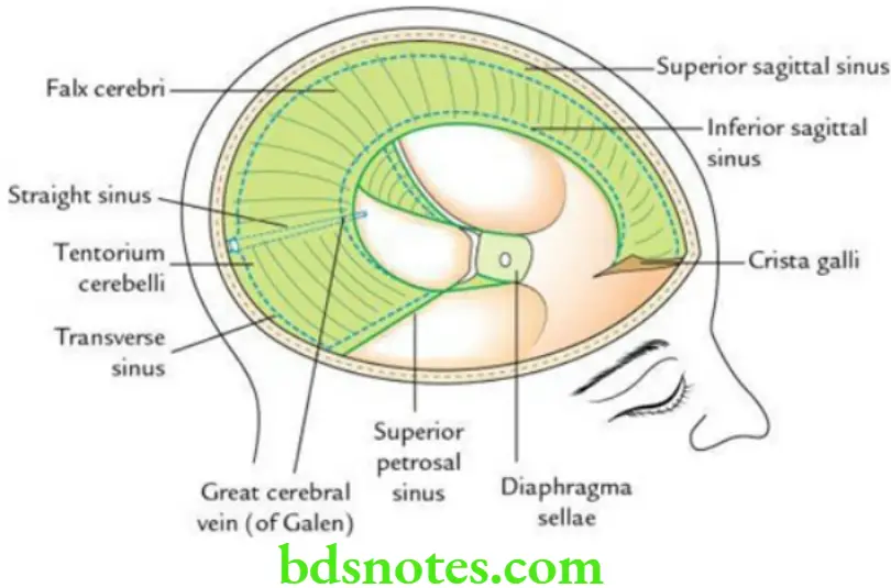
The dural folds are generally formed by folding of the meningeal layer of the cranial dura, which projects into the cranial cavity to divide it into various compartments, which lodge the different lobes/parts of the brain.
The folds of dura mater are as follows:
- Falx cerebri
- Tentorium cerebelli
- Falx cerebelli
- Diaphragma seller
Question 2. Briefly describe falx cerebri.
Answer.
Falx Cerebri is a large, sickle-shaped fold of dura mater that lies in the median longitudinal fissure between the two cerebral hemispheres.
Falx Cerebri Features It presents two ends – anterior and posterior; two margins – upper and lower; and two surfaces – right and left.
Falx Cerebri Anterior end: It is narrow and attached to the frontal crest of the frontal bone and crista galli of the ethmoid bone.
Falx Cerebri Posterior end: It is broad and attached along the median plane to the upper surface of the tentorium cerebelli.
Falx Cerebri Upper attached margin: It is convex and attached to the lips of the sagittal sulcus on the inner aspect of the cranial vault as far back as the internal occipital protuberance.
Falx Cerebri Lower free margin: It is concave, free and lies close to the corpus callosum.
Falx Cerebri Right and left surfaces: Each of these surfaces is related to the medial surface of the corresponding cerebral hemisphere.
Venous sinus enclosed in the falx cerebri
- Superior sagittal sinus along the upper margin
- Inferior sagittal sinus along the lower margin
- Straight sinus along the line of attachment of the falx cerebri to the tentorium cerebelli
Falx Cerebri Applied anatomy
Thrombosis of the superior sagittal sinus may occur due to the spread of infection into it from the nose, scalp and diploic veins. It interferes with the absorption of CSF, leading to increased intracranial pressure.
Question 3. Briefly describe Tentorium Cerebelli.
Answer.
Tentorium cerebelli is a tent-shaped fold of dura mater, which forms the roof of the posterior cranial fossa. It separates the cerebellum from the occipital lobes of the cerebrum.
Tentorium Cerebelli Features It has two margins and two surfaces.
Tentorium Cerebelli Margins
- The inner free margin is U-shaped and encloses the tentorial notch for the passage of the midbrain. The anterior ends of the concave free margin are attached to the anterior clinoid processes.
- The outer attached margin is convex and attached on each side (from anterior to posterior) to the posterior clinoid process, superior border of petrous temporal bone, posteroinferior angle of parietal bone and lips of transverse sulcus on occipital bone.
Tentorium Cerebelli Surfaces
- The convex upper surface slopes on either side from the median plane. In the median plane, it provides attachment to falx cerebri.
- The concave inferior surface provides attachment to the falx cerebelli in its posterior part.
Venous sinus enclosed in tentorium cerebelli
- Superior petrosal sinus within the anterolateral part of the attached margin.
- Transverse sinus within the posterior part of the attached margin.
- Straight sinus along the line of attachment between falx cerebri and tentorium cerebelli.
Question 4. Define Intracranial Dural Venous Sinuses. Discuss their characteristic features and applied anatomy.
Answer.
Intracranial Dural Venous Sinuses Definition The intracranial dural venous sinuses are endothelial-lined venous channels lying between the two layers of the dura mater.
Intracranial Dural Venous Sinuses Characteristic features
- Are devoid of smooth muscle in their wall.
- Are devoid of valves in their lumen.
- Are lined by endothelium.
- Drain CSF through arachnoid villi and granulations.
Read And Learn More: Selective Anatomy Notes And Question And Answers
- Receive emissary veins.
- Communicate with extracranial veins through emissary veins.
- Communicate with vertebral venous plexus through basilar venous plexus.
- Receive veins from the brain and diploic veins from cranial bones.
- Noncompressive in nature (i.e. always kept patent) and equalize pressure within and outside the skull.
Intracranial Dural Venous Sinuses Applied anatomy The infection from extracranial sources can spread to the dural venous sinuses and then to the brain.
Question 5. Write about the classification of intracranial dural venous sinuses.
Answer.
The intracranial dural venous sinuses are classified into two types – paired and unpaired.
Classification of Intracranial Dural Venous Sinuses
Question 6. Describe the Cavernous Sinus under the following headings:
- Formation and location,
- Relations,
- Contents,
- Tributaries and communications and
- Applied anatomy.
Answer.
Cavernous Sinus Formation and location
The two cavernous sinuses are situated on either side of the pituitary fossa and the body of the sphenoid. Each cavernous is a large venous space (2 cm long and 1 cm wide) formed by the separation of endosteal and meningeal layers of dura mater, lined by endothelium. Its floor is formed by the endosteal layer, whereas its lateral wall, roof and medial wall are formed by a meningeal layer of the dura mater.
Cavernous Sinus Relations
Cavernous Sinus Superior: Optic chiasma, internal carotid artery and anterior perforated substance.
Cavernous Sinus Inferior: Foramen lacerum and greater wing of sphenoid.
Cavernous Sinus Medial: Hypophysis cerebri and sphenoidal air sinus.
Cavernous Sinus Lateral: Temporal lobe of the brain (uncus) and cavum trigeminal with trigeminal ganglion within it.
Cavernous Sinus Contents
Structures present in the lateral wall: From anterior to posterior, these are
- Oculomotor nerve (CN 3)
- Trochlear nerve (CN 4)
- Ophthalmic nerve (CN V1)
- Maxillary nerve (CN V2)
Cavernous Sinus Structures passing through the sinus
- An internal carotid artery with a sympathetic plexus around it
- Abducent nerve – below and lateral to the internal carotid artery
Cavernous Sinus Tributaries
Cavernous Sinus Communications
- The superior petrosal sinus connects it with the transverse sinus.
- The inferior petrosal sinus connects it with the internal jugular vein.
- Emissary veins connect it with the pterygoid venous plexus.
- The ophthalmic vein connects it with the facial vein.
- Anterior and posterior intercavernous sinuses connect it with the opposite cavernous sinus.
Cavernous Sinus Applied anatomy
- Thrombosis of Cavernous Sinus: It may be caused by the spread of septic infection in the dangerous area of the face.
The clinical features of cavernous sinus thrombosis are as follows:- Severe pain in the eye due to involvement of the ophthalmic nerve.
- Ophthalmoplegia due to involvement of CN 3, 4 and 6.
- Oedema of eyelids due to congestion of orbital veins.
- Exophthalmos due to congestion of orbital veins.
- Arteriovenous Fistula: It is caused by a rupture of the internal carotid artery into the cavernous sinus. It results in:
- Unilateral pulsating exophthalmos
- Loud systolic thrill/murmur over the eye
Question 7. Define emissary veins. Enumerate the important emissary veins and discuss their functional and clinical importance.
Answer.
The emissary veins are thin-walled venous channels that connect the extracranial veins to the intracranial dural venous sinuses.
Following are the important emissary veins:
- Parietal emissary vein
- Mastoid emissary vein
- An emissary vein passing through the foramen ovale or emissary sphenoidal foramen
- Small emissary veins pass through the foramen lacerum and connect the cavernous sinus to the pterygoid plexus of veins
- An emissary vein passes through the posterior condylar canal and connects the sigmoid sinus to the suboccipital plexus of veins
- Ophthalmic veins can also act as emissary veins
Emissary Veins Functions The emissary’s veins help to maintain the constant intracranial pressure because blood passing through them can flow in either direction according to the state of intracranial pressure.
Emissary Veins Functions Clinical importance They can carry infection from outside the skull to inside the skull (i.e. the cranial cavity).
Question 7. Describe the pituitary gland under the following headings:
- Definition and location,
- Gross features,
- Relations,
- Microscopic structure,
- Blood supply,
- Nerve supply,
- Development and
- Applied anatomy.
Answer.
Definition and location Pituitary Gland: The pituitary gland/hypophysis cerebri is a small neuroendocrine gland located in the hypophyseal fossa (sella turcica) of the body of the sphenoid.
Pituitary Gland Shape and Measurements
Pituitary Gland Gross features
The pituitary gland consists of two parts, which are embryologically, morphologically and functionally different from each other:
- Adenohypophysis (anterior pituitary)
- Neurohypophysis (posterior pituitary)
Adenohypophysis (anterior pituitary):
It is highly cellular and presents an intraglandular cleft. It is further subdivided into three parts/lobes:
- Anterior lobe (pars distalis): It is the major part of the adenohypophysis.
- Intermediate lobe (pars intermedia): It is a thin strip of glandular tissue between the intraglandular cleft in front and neurohypophysis behind.
- Tuberal lobe (pars tuberalis): It is an upward extension of the anterior lobe that surrounds the part of the infundibulum.
Neurohypophysis (posterior pituitary):
It is continuous above the infundibulum, which extends downward and forward from the floor of the 3rd ventricle and enters the hypophyseal fossa after piercing the diaphragm sellae. It is subdivided into three parts:
- Posterior lobe (pars posterior): It is smaller than the anterior lobe and lies in the posterior concavity of the larger anterior lobe.
- Infundibulum (neural stalk): It contains the neural connections of the neurohypophysis.
- Median eminence of the tuber cinereum: It is continuous with the infundibular stem.
Pituitary Gland Relations
Anterior:
- Anterior intercavernous sinus
Posterior:
- Posterior intercavernous sinus
- Dorsum sellae
- Basilar artery
- Pons
Superior:
- Diaphragma sellae
- Optic chiasma
- Tuber cinereum
- Infundibular recess of the 3rd ventricle
Inferior:
- Hypophyseal fossa
- Body of the sphenoid
- Sphenoidal air sinuses
Lateral (on each side):
- Cavernous sinus with its contents
Pituitary Gland Microscopic structure
Anterior lobe (pars anterior/distal): It forms three-fourths of the gland. It consists of two types of glandular cells arranged in irregular cords or clumps:
- Chromophilic cells (50%) having affinity to colours, i.e. staining
- Acidophils/alpha cells (about 43% of cells): Secrete GH, ACTH and prolactin
- Basophils/beta cells (about 7% of cells): Secrete TSH, LH, FSH and MSH
- Chromophobic cells (50%), which do not take the colour of stain: Resting cells, no secretion
Intermediate lobe (pars intermedia): It is made up of numerous basophilic and chromophobic cells. The cells are arranged in the form of small follicles containing colloid material.
Posterior lobe (pars posterior/nervosa): It consists of:
- A large number of nonmyelinated nerve fibres.
- Modified neuroglial cells called pituicytes.
- Presence of Herring bodies, the small, spherical masses that stain deeply with chrome–alum haematoxylin.
Pituitary Gland Arterial supply:
Venous drainage: Small short veins emerge from the surface of the gland and drain into neighbouring dural venous sinuses.
Pituitary Gland Nerve supply: By hypothalamo-hypophyseal tract, which arises from preoptic and paraventricular nuclei of the hypothalamus.
Pituitary Gland Development: The pituitary gland develops from two distinct sources:
- Adenohypophysis develops from Rathke’s pouch – an ectodermal diverticulum (outpocketing) from the roof of the stomodeum that grows cranially in front of the buccopharyngeal membrane.
- Neurohypophysis develops from the infundibulum – a down growth from the floor of 3rd ventricle.
Pituitary Gland Applied Anatomy The pituitary gland produces a number of hormones that control the secretions of other endocrine glands of the body.
- Pituitary adenoma: It is not uncommon. It compresses the central part of optic chiasma leading to bitemporal hemianopia (tunnel vision).
- Gigantism and acromegaly: Gigantism occurs due to excessive secretion of growth hormone (GH) before adolescence; hence, the person becomes very tall (8–9 feet) due to excessive length of the bone. The acromegaly occurs due to excessive secretion of GH in adults leading to coarse facial features with protrusion of jaw (prognathism).
- Pituitary dwarfism: Occurs due to hyposecretion of GH causing shortening of limbs.
Leave a Reply