Dura Mater
Question 1. Duramater
Answer:
- It is the outermost, thickest & toughest membrane covering the brain
Layers:
1. Outer / Endosteal layer
- Serves as endosteum for the skull bones
- It is richly vascular
2. Inner/Meningeal layer
- Surrounds the brain
- It is folded to divide the cranial cavity into compartments
- It is more fibrous & requires little blood supply
“Understanding the dura mater through FAQs: Anatomy, functions, and uses explained”
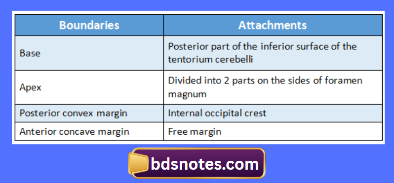
Question 2. Falx cerebelli
Answer:
- It is small sickle shaped fold of duramater
- It projects into the posterior cerebellar notch
“Importance of studying the dura mater for medical students: Questions explained”
Question 3. Straight sinus
Answer:
- It lies within the junction of the falx cerebri & the tentorium sellae
- It is formed by the union of the inferior Sagittal sinus with the great cerebral vein
- Here exists a ball valve mechanism formed by sinusoidal plexus of blood vessels
- This regulates the secretions of CSF
- It ends at the internal occipital protuberence
Question 4. Transverse sinus
Answer:
- They are paired large sinuses
- They are situated in the posterior part of the attached margin of the tentorium cerebelli
- The right transverse sinus is continuation of the superior sagittal sinus & left sinus is continuation of the straight sinus
“Common challenges in mastering dura mater notes effectively: FAQs provided”
Extend:
- From the internal occipital protuberence to the posteroinferior angle of the parietal bone
Tributaries:
- Diploic vein
- Superior petrosal sinus
- Inferior cerebral vein
- Inferior cerebellar vein
- Inferior anastomic vein
“Factors influencing success with dura mater studies: Q&A”
Question 5. Sigmoid sinus
Answer:
- It is paired S-shaped sinus
- It is direct continuation of the transverse sinus
Extend:
- From the posteroinferior angle of the parietal bone to the posterior part of the jugular foramen
Tributaries:
- Mastoid & Condylar emissary vein
- Cerebellar vein
- Internal auditory vein
Leave a Reply