Diseases Of The Pulp And Periapical Tissues
Question.1. Describe the etiology, histopathology, and clinical features of acute pulpitis.
Or
Describe etiology, histopathology, clinical features, and sequelae of acute pulpitis.
Answer. Acute pulpitis is an irreversible condition characterized by acute, intense inflammatory reaction in pulp tissue.
Etiology
- Pulp exposure due to faulty cavity preparation.
- Caries progressing beyond the dentinal barriers and reaching the pulp.
- Chemical irritation to pulp
- Cracked tooth syndrome
- Metallic restoration in a tooth without proper thermal insulation.
- Blow to tooth with subsequent damage to pulp.
- Recurrent caries around the preexisting restoration.
- Galvanic current produced due to dissimilar metallic restoration may transmitted to pulp and causing pulpitis.
Read And Learn More: Oral Pathology Question And Answers
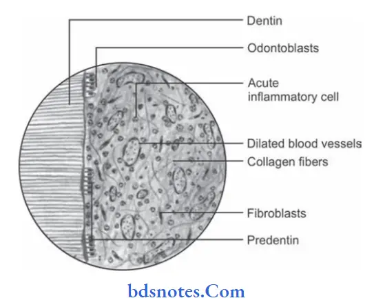
Histopathology
- Severe edema in the pulp with vasodilatation.
- Moderate to dense infiltration of polymorphonuclear leukocytes.
- Focal of complete destruction of odontoblast cells at pulp dentin border.
- Many micro abscess formations in pulp characterized by the area of liquefaction degeneration in pulp being surrounded by dense band of neutrophils and microorganisms.
- There may be complete liquefaction and necrosis of pulp with total destruction of odontoblastic cell layer. This is known as acute suppurative pulpitis.
- Death of pulp is due to tissue dehydration. This is known as “dry gangrene of pulp”.
Clinical Features
- Tooth is sensitive to cold and hot stimuli.
- Application of hot or cold stimuli causes an increase in intensity to pain and such pain persists for longer duration even after the stimuli is removed.
- Intensity of pain increases during the sleep and occurs due to increase in local blood pressure in head and neck region.
- As entrance of pulp is not wide, acute pulpitis helps in spread of inflammation throughout pulp with subsequent necrosis.
- Acute pulpitis is often associated with microabscess formation in pulp along with liquefaction degeneration.
Pain subsides when drainage is established or when pulp undergoes complete necrosis. - Tooth is nontender to percussion unless the pulpal inflammation has spread beyond the root apex into periapical region.
- When intrapulpal pressure becomes very high during acute inflammation it cause collapse of apical blood vessels. This is known as “pulp strangulation”.
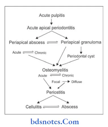
Sequelae Of Acute Pulpitis
Question.2.What is pulpitis? Explain diffrent causes of pulpitis.
Describe clinical features and histopathological picture of chronic open hyperplastic pulpitis.
Answer. Pulpitis refers to the inflammation of dental pulp within the tooth.
Causes of pulpitis
- Reversible pulpitis: It is caused by an agent capable of injuring the pulp like trauma, disturbed occlusal relationship and thermal shock.
- Irreversible pulpitis: It is caused by the bacterial involvement of pulp through caries, chemical or thermal or mechanical injury.
Chronic open hyperplastic pulpitis
- It is also known as chronic hyperplastic pulpitis or pulp polyp or pulpitis aperta.
- It is a productive pulpal inflammation due to an extensive carious exposure of young pulp. It is characterized by development of granulation tissue covered by epithelium and resulting from long-standing low-grade infection.
Clinical Features
- Pulp polyp appears as small, pinkish, red-lobulated mass, which protrudes from pulp chamber and fills up the carious cavity.
- Condition is seen in young adults and children. It commonly develops in deciduous molar and first permanent molars.
- The affected tooth has a large open carious cavity, which is present for long duration.
- Lesion bleeds profusely on provocation.
- Involved tooth is painless and is sensitive to thermal stimuli.
Histopathology
- Hyperplastic pulp tissue lesion presents the feature of granulation tissue mass, consisting of numerous proliferating firoblasts and young blood capillaries.
- Inflammatory cell infiltration by lymphocytes, plasma cells and sometimes polymorphonuclear neutrophils in tissue are common.
- Stratified squamous epithelium is present on the surface of hyperplastic pulpitis which resembles oral epithelium.
- Epithelium surface show well-formed rete peg formation.
- Epithelial cells on surface are believed to be desquamated epithelial cells which came either from buccal mucosa or from salivary gland ducts.
Question.3. Write short note on pulpitis.
Answer.
Chronic pulpitis
It is a condition characterized by the low-grade fever often persistent inflammatory reaction in pulpal tissue with litte or no constitutional symptoms.
Clinical Features
- The tooth with chronic pulpitis may remain asymptomatic for long time.
- There may be an intermittent dull and throbbing pain in the tooth.
- Tooth is less sensitive to hot and cold stimuli.
- Tooth responds to a higher level of current when electric pulp tester is used.
- Exposed pulp tissue may be manipulated by small instrument but bleeding can occur.
Histopathology
- Chronic inflammatory response in the pulp is characterized by cellular infiltration by the lymphocytes, plasma cells, and macrophages.
- Prolong chronic inflammation may encourage fibroblast activity in pulp with formation of collagen bundle and in some cases leads to internal resorption of the tooth.
- Blood capillaries are prominent and few microorganisms are found in the pulpal tissue.
- Persisting chronic pulpitis may cause diffuse or solitary areas of calcification in the pulp.
Treatment
- Extraction of tooth
- Root canal therapy
Question.4. Write short note on pulp polyp.
Or
Write short note on chronic hyperplastic pulpitis.
Or
Write short answer on pulp polyp.
Answer.
Treatment
- Elimination of polypoid tissue, following the extirpation of the pulp.
- After removing hyperplastic pulp tissue bleeding can be stopped by pressure.
- Extraction of tooth or root canal treatment.
Question.5. Describe etiology, histopathology, and clinical feature of periapical granuloma.
Answer. It is also called as chronic apical periodontitis.
Periapical granuloma is a localized mass of granulation tissue around the root apex of nonvital tooth which develop in relation to infection and inflammation.
Etiology
- Extension of pulpal inflammation
- Occlusal trauma
- Orthodontic tooth movements with excessive uncontrolled force
- Acute trauma due to blows on tooth.
- Spread of periodontal infection in root apex.
- Perforation of root apex into endodontic therapy.
Clinical Features
- Tooth involves produce sensitivity to percussion which occurs due to edema, hyperemia and inflammation of apical periodontal ligament.
- Mild pain and discomfort in tooth during chewing solid foods.
- Involved tooth is slightly elongated from the socket.
- Periapical granuloma may be asymptomatic in many cases.
- Tooth may be vital or partially vital in initial stages of development of lesion but in fully developed periapical granuloma the affected tooth is nonvital.
Histopathology
- Lesion appears as granulation tissue mass consisting of proliferating fibroblasts, endothelial cells and numerous immature blood capillaries.
- Chronic inflammatory cells, i.e. macrophages, lymphocytes, and plasma cells are present in the lesion.
- There is presence of epithelial islands, cholesterol clefts, and foam cells.
- Plasma cells often produce immunoglobulin there is also present of T lymphocytes in the lesion.
- Epithelial rest cell of malassez, proliferate in response to chronic inflammation and these proliferating cells undergo certification.
- Bony tissue at the periphery of lesion is lined by the osteoclast cells with area of bone resorption.
- Few bacterias are present in the lesions which are not affected by the cellular immune mechanism.
- Occasionally Russell bodies are also found.
- Resorption of cementum and dentin often occurs as a result
of chronic inflammation. In some areas along root, cemento
blastic activity predominates leading to hypercementosis.
Question.6. Describe etiology, histopathology and clinical features of acute alveolar abscess.
Answer. It is also known as dentoalveolar abscess.
It is defined as acute suppurative infection in periapical region of tooth.
Etiology
- Extension of pulpal infection in periapical tissue.
- Fracture of tooth with pulp exposure.
- Accidental perforation of apical foramen during root canal treatment
- Extension of periodontal infection in periapical tissues.
- Anachoretic infection of periapical tissues.
Clinical Features
- It is common odontogenic infection and constitutes 2% of apical radiolucencies.
- Due to acute abscess there is pain in the affcted tooth.
- Localized swelling and an erythematous change in overlying mucosa is present.
- Affected area of jaw may be tendered during palpitation.
- Pain aggravates during percussion and when pressure is applied with the opposing tooth.
- Application of heat intensifies pain, whereas application of cold relieves pain temporarily.
- Pus discharging sinus often develops on alveolar mucosa over the affected root apex and sometimes on skin overlying the jaw bone.
- Infection from acute periapical abscess often spreads to facial spaces, leading to space infections.
Histopathology
- Lesion appears as zone of liquefaction necrosis, which is made up of proteinaceous exudates, necrotic tissue and large number of dead neutrophils
- Adjacent tissue surrounding the bone has many dilated blood vessels and infitration with the neutrophils.
- Inflammatory changes are observed in the PDL and adjoining bone marrow.
- Bony trabeculae in peri apical region may show empty lacunae, which results from death of osteocytes.
Question.7. Describe the etiology, histopathology and clinical features of acute suppurative osteomyelitis in adult patient mandible.
Or
Describe etiology, histopathology and clinical features and complications of acute suppurative osteomyelitis in adult patient mandible.
Answer. Acute suppurative osteomyelitis is serious sequelae of periapical infection, there is diffuse spread of infection throughout medullary spaces with subsequent necrosis of variable amount of bone.
Etiology
- Direct spread of infection from dental pulp into the mandible.
- Spread of infection in the mandible from presenting suppurative odontogenic infections.
- Spread of infection following removable of tooth without proper asepsis and antibiotic coverage.
- Compound fracture of mandible with exposure of bone outside the mucosa.
- Postradiation secondary infection.
- Infection to the preexisting bony lesions, e.g. Paget’s disease of bone and firous dysplasia.
Clinical Features
- It occurs after the 50 years of age and males are more commonly affected.
- Mandibular lesions are diffuse in nature.
- Acute suppurative osteomyelitis of mandible in young adult causes severe pain, and diffuse and enlarged swelling of mandible.
- There is loosening and soreness of the regional teeth with difficulty in food intake.
- Multiple intraoral and extraoral pus-discharging sinuses often develops and moreover discharge of pus is seen from gingival cervice of the affected teeth.
- Paresthesia of lip is common.
- Patient is slightly febrile and general symptoms include fever, malaise, anorexia and vomiting.
Histopathology
- In acute suppurative osteomyelitis bone marrow undergoes liquefaction and purulent exudates occupy the marrow space.
- A large number of acute inflammatory cells infiltrations are present which shows PMNs with occasional presence of lymphocytes and plasma cells.
- Some areas of affected bone undergo necrosis with generation of osteoblast and osteocytes cells and therefore results in development of sequestrum (a piece of dead bone).
- When acute phase of infection subsides in new shell of bone called “involucrum” is formed over inflammatory focus.
Complications
- Periostitis
- Cellulitis
- Abscess.
Question.8.Defie and classify osteomyelitis. Describe in detail chronic osteomyelitis.
Or
Write short note on chronic osteomyelitis.
Answer. Chronic osteomyelitis is the persistent abscess of the bone characterized by the complex inflammatory process including necrosis of mineralized and marrow tissues, suppuration, resorption, sclerosis, and hyperplasia.
Osteomyelitis is defined as an inflammatory condition of bone that begins as an infection of medullary cavity and haversian systems of the cortex and extends to involve the periosteum of the affected area.
Classification of Osteomyelitis
- Acute osteomyelitis
- Acute suppurative osteomyelitis
- Acute subperiosteal osteomyelitis
- Acute periostitis
- Chronic osteomyelitis
- Non-Specific Type
- Chronic intramedullary osteomyelitis
- Chronic focal sclerosing osteomyelitis
- Chronic diffuse sclerosing osteomyelitis
- Chronic osteomyelitis with proliferative periostitis
- Chronic subperiosteal osteomyelitis
- Chronic periostitis
- Specific type
- Tuberculous osteomyelitis
- Syphilitic osteomyelitis
- Actinomycotic osteomyelitis
- Non-Specific Type
- Radiation-induced osteomyelitis
- Idiopathic osteomyelitis
Pathogenesis
Clinical Features
- Molar area of mandible is more frequently affected.
- Pain is usually mild and insidious and is not related to the real severity of disease.
- Jaw swelling is common feature but mobility of teeth and sinus tract formation are rare.
- Anesthesia and paresthesia of lip is uncommon.
- Regional lymphadenopathy is common.
- There is thickened, woodened feeling of bone and slow increase in jaw size.
Histopathology
- Chronic inflammatory reaction of bone with accumulation of exudate and pus within medullary spaces.
- Lymphocytes, macrophages, and plasma cells predominate among the inflammatory cells.
- Osteoblastic and osteoclastic cavity occurs partially with formation of irregular bony trabeculae having reversal lines.
- Sequestrum may develop in later stages of the disease.
Colonies of bacteria are also seen within the inflmed tissue.
Question.9.Classify pulpitis and write its sequelae. Write in short etiology, clinical features, roentgenographic features,histology with treatment and prognosis of Garre’s osteomyelitis.
Answer. Inflammation of pulp is called as pulpitis.
Classifiation of Pulpitis
Reversible
- Symptomatic (acute)
- Asymptomatic (chronic).
Irreversible
- Acute – Abnormally responsive to cold
- Abnormally responsive to heat
- Chronic – Asymptomatic with pulp exposure
- Hyperplastic pulpitis
- Internal resorption.
Garre’S Osteomyelitis
- It is also called as chronic osteomyelitis with proliferative periostitis or periostitis ossifies or Garre’s chronic non-suppurative sclerosing ostitis.
- Garre’s osteomyelitis represents a reactive periosteal osteogenesis in response to low grade infection or trauma.
Etiology
- Mild infection
- Chronic periapical abscess
- Infected periapical cyst
- Mechanical irritation in the jaw from dentures
- Chronic trauma in the jaw bone.
Sequelae Of Pulpitis
Clinical Features
- It is common in young children and adults.
- Mandible is commonly involved in the posterior part.
- The involved jaw bone presents a carious nonvital tooth.
- There is a slight tenderness or a vague pain may be felt in the affected area of the bone.
- Slight pyrexia and leukocytosis may be present but ESR is normal.
Roentgenographic Features
- There is presence of a shadow of thin convex shell of bone over cortex.
- As the infection proceeds cortex become thick and laminated with alternating radiolucent and radiopaque layers. This is also known as onion skin appearance.
- Cancellous bone adjacent to the lesion can be normal, become sclerotic or it can show some areas of osteolytic changes.
- In the new bone osteolytic radiolucencies, i.e. small sequestra are seen.
Histology
- There is presence of newly formed bone consisting of multiple osteoids and primitive bony tissue in sub periosteal region.
- Osteoblastic as well as osteoclastic activities are observed in central part of the bone.
- Marrow space contains firous tissues showing patchy areas of chronic inflammatory cell infiltration
- Trabeculae are oriented perpendicular to the cortex with trabeculae arranged, parallel to each other shows “retiform pattern”.
- Connective tissue between the bony trabeculae shows a diffuse or patchy sprinkling of lymphocytes and plasma cells.
Treatment And Prognosis
- Elimination of causative agent
- Extraction of carious infected tooth and antibiotic therapy
- Prognosis is good so no any additional surgical intervention is required.
Question.10. Write short note on phoenix abscess.
Answer. Acute exacerbation of chronic periapical lesion is called as phoenix abscess.
Phoenix abscess is also known as recrudescent abscess.
Phoenix abscess is also defined as an acute inflammatory reaction superimposed on existing chronic lesion such as cyst or granuloma.
Etiology
Chronic periradicular lesions such as granulomas are in state of equilibrium during which they can be completely asymptomatic.
But, sometimes, influx of necrotic products from diseased pulp or bacteria and their toxins can cause the dormant lesion to react and this leads to initiation of acute inflammatory response.
Lowered body defenses and mechanical irritation during root canal treatment also trigger an acute inflammatory response.
Clinical features
- First symptom is that, there is tenderness on percussion.
- Tooth is slightly extruded from socket and is mobile.
- Patient can or cannot have swelling. Swelling, if present is localized and if left untreated may become diffuse (cellulitis), leading to asymmetry of the patients face. In case of upper canines, it may even extend to the eyelids.
- Patient can be present with fever, malaise and lymphadenopathy.
- Tissue at the surface of swelling appears taut and inflamed;pus starts to form beneath it.
- As the liquefaction continues, tissue ruptures due to the pressure to form a sinus tract which opens on the labial/buccal mucosa. This process is the beginning of chronic alveolar abscess.
Histopathology
There is presence of areas of liquefaction necrosis with disintegrated polymorphonuclear leukocytes and cellular debris surrounded by macrophages. lymphocytes, plasma cells in periradicular tissues.
Treatment
- Establishment of drainage is done.
- As symptoms subsides root canal therapy should be done.
Question.11. Describe in brief etiology of pulpitis.
Answer. Following is the etiology of pulpitis
- Dental caries which extend beyond the dentinal barrier and reaches pulp lead to pulpitis.
- During cavity preparation if pulp exposure occurs this will lead to pulpitis.
- When blow to the tooth occur which lead to damage of pulp.
- If cavity preparation is done without using the water spray this lead to the excessive heat production to tooth which lead to pulpitis.
- Chemical irritation to pulp
- Cracked tooth syndrome
- If metallic restoration is given in the tooth without providing proper thermal insulation this will lead to pulpitis.
Question.12. Write short note on periapical abscess.
Answer. Periapical abscess is an acute or chronic suppurative process of dental periapical region.
It is also known as dentoalveolar abscess or alveolar abscess.
Acute exacerbation of chronic periapical lesion is called as phoenix abscess.
Types Of Periapical Abscess
Acute: It is associated with severe pain in tooth.
Chronic: It is long standing and symptoms are of low grade.
Clinical Features
Acute Periapical Abscess
- Patient complains of severe pain which is of throbbing variety.
- There is also presence of swelling in the associated area.
- Mucosa surrounding the swelling becomes tough and inflmed.
- Slight fever is present.
- Regional lymphadenitis is present.
- Patient feels sensitivity with the affcted tooth.
- Tooth is tender to palpation and is mobile.
Chronic periapical abscess
- Pain is present from a longer time. Nature of pain is dull.
- Sinus formation is seen either intraorally and extraorally.
- At opening of sinus mass of inflmed granulation tissue is present known as parulis.
- Lymphadenopathy is present.
Histopathology
- Area of suppuration consists of central area of disintegrating PMN leukocytes surrounded by lymphocytes, cellular debris, necrotic material and the bacterial colonies.
- Dilated blood vessels are seen in PDL and adjacent marrow spaces of bone.
- Inflammatory cell infiltrate is seen in marrow spaces.
- In chronic periapical abscess chronic inflammatory infitrate consist of lymphocytes, plasma cells, macrophages.
Treatment
Drainage of abscess is done followed by opening of root canal or extraction of tooth.
Question.13. Describe etiopathogenesis of periapical granuloma.
Answer. Following is the etiopathogenesis of periapical granuloma:
- Periapical granuloma is caused as a response to prolonged irritation from infected root canals which leads to the extension of chronic apical periodontitis in PDL.
- Since pulp is infected it presents the release of inflammatory mediators such as prostaglandins,kinins and endotoxins. Elevated levels of IgG are seen in pulpoperiapical lesion.
- Inflammation as well as increase in the vascular pressure leads to abscess formation and resorption of bone in affcted area which is replaced by the granulation tissue.
Leave a Reply