• Skip to main content
  • Skip to secondary menu
  • Skip to primary sidebar
  • About Us
  • Terms of Use
  • Privacy Policy
  • Disclaimer
  • Contact Us
  • Sitemap

BDS Notes

BDS notes, Question and Answers

  • Public Health Dentistry
  • Periodontics
  • Pharmacology
  • Pathology
  • Orthodontics
    • Anchorage In Orthodontics
    • Mandibular Growth, Functional Matrix
    • Retention and Relapse
  • General Surgery
    • Cysts: Types, Causes, Symptoms
    • Maxillofacial Fractures, Disorders, and Treatments
    • Lymphatic Disorders
    • Neurological and Facial Disorders
  • Temporal And Infratemporal Regions
    • Spinal and Neuroanatomy
  • Dental Materials
    • Dental Amalgam
Home » Difference Between Upper And Lower Motor Neurons

Difference Between Upper And Lower Motor Neurons

February 6, 2026 by Kristensmith Taylor Leave a Comment

Difference Between Upper And Lower Motor Neurons

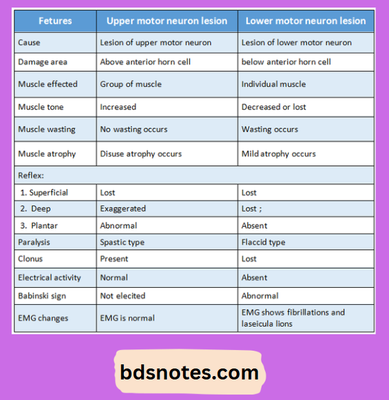
Question 1. State the difference between upper motor neuron and lower motor neuron lesions.
Answer:

Upper Lower Motor Neuron Lesions Comparison

Question 2. Functional unit of nervous system.
Answer:

Neuron:

  • The neuron is made up of.

1. Nerve cell body/soma:

  • It is irregular in shape.
  • It constitutes of
    1. Cytoplasm called neuroplasm. It contains.
      1. Nucleus.
        • Large nucleus covers central part of the nerve cell body and has two or more nucleoli.
      2. Nissl granules/bodies.
        • They are composed by many thin, parallely arranged, membrane bounded cavities.
        • They are covered by minute particles containing RNA with proteins.
      3. Neurofibrils:
        • They are thread like structures consisting of microfilaments and microtubules.
      4. Mitochondria and golgi apparatus.
    2. Cell membrane – covers neuroplasm:

2. Dendrite:

  • It is the branched process of the neuron.
  • It transmits impulses towards nerve cell body.
  • It contains nissl granules, mitochondira and neurofibrillae.
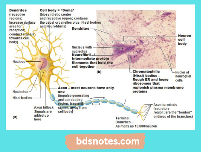
Spinal Motor Neuron Structure

3. Axon:

  • It originates from thickened area of the cell body called Avon Hillock.
  • It is the longer process of the nerve cell.
  • It conducts impulses away from the cell body
  • The cytoplasmic fluid occupying the centre of the axon is called axoplasm.
  • The cell membrane covering it is called axolemma.
  • A short distance from its origin, the axon acquires myelin sheath.
  • Axoplasm contains mitochondira, neurofibrils and axoplasmic vesicles but nissl bodies are absent.

Filed Under: Anatomy

Reader Interactions

Leave a Reply Cancel reply

Your email address will not be published. Required fields are marked *

Primary Sidebar

Recent Posts

  • Branchial Cleft Cyst: Background, Pathophysiology, Etiology
  • Maxillary Nerve: Origin, Course, And Branches
  • The Father Of Anatomy And A Great Anatomist Herophilus
  • Bone Structure – Anatomy
  • The External Carotid Artery: Anatomy, Branches, And Functions
  • Occipitofrontalis Muscle
  • Superficial Temporal Artery
  • Platysma Muscle
  • Cartilage
  • Cauda Equina And Conus Medullaris Syndromes
  • Subcutaneous Injections And Device Management
  • Types Of Circulation: Pulmonary, Systemic, And Portal
  • Structure Of Skeletal Muscle
  • Elastic Cartilage
  • Cellular Organelles And Structure
  • The Golgi Apparatus – The Cell
  • The Cytoplasmic Inclusions Of Certain Plant Cells
  • Dental Abscess
  • Laser Surgery
  • Our Facial Muscles And Their Functions

Copyright © 2026 · Magazine Pro on Genesis Framework · WordPress · Log in